
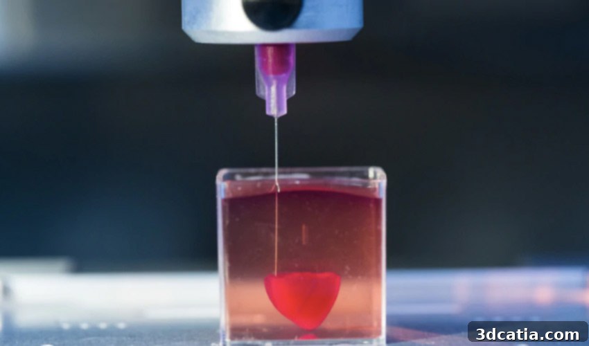
Revolutionary Bioprinting: Tel Aviv University Scientists Unveil World’s First 3D Printed Vascularized Heart from Human Tissue
In a monumental stride for medical science and regenerative medicine, researchers at Tel Aviv University have achieved an unprecedented feat: the successful 3D printing of a small, vascularized heart using human tissue. This groundbreaking achievement marks the first time a fully cellularized and vascularized heart, complete with blood vessels, ventricles, and chambers, has been engineered and printed. What makes this breakthrough even more remarkable is that the printed heart perfectly matches the immunological, cellular, and anatomical properties of a human patient, significantly reducing the risk of rejection upon transplantation. Published on April 15th in a pivotal study in *Advanced Science*, these findings open up transformative possibilities for personalized organ replacement and the future of healthcare. Until now, scientists had only managed to print simpler tissues without the complex network of blood vessels, which are essential for nutrient supply and waste removal in larger organs.
Professor Tal Dvir of TAU’s School of Molecular Cell Biology and Biotechnology, who spearheaded this pioneering research, emphasized the significance of their accomplishment. “This is the first time anyone anywhere has successfully engineered and printed an entire heart replete with cells, blood vessels, ventricles, and chambers,” stated Prof. Dvir. This innovation comes at a critical time, as heart disease continues to be the leading cause of death among both men and women globally. For patients suffering from end-stage heart failure, heart transplantation remains the only viable treatment option. However, the severe and ever-growing shortage of donor hearts presents a profound challenge, highlighting an urgent need for alternative approaches to repair or replace diseased hearts.
The scarcity of donor organs is a global health crisis, with thousands of patients dying each year while awaiting a suitable transplant. Traditional organ transplantation carries inherent risks, including the potential for immune rejection and the lifelong need for immunosuppressive drugs, which can have significant side effects. The ability to bio-print patient-specific organs could revolutionize transplant medicine by eliminating the need for donor matching and reducing the risks associated with immune response. This development offers a beacon of hope for countless individuals whose lives are currently dependent on the availability of a matching donor.
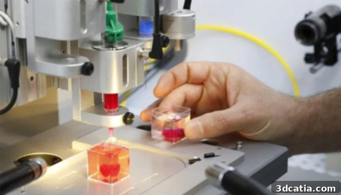
Central to this breakthrough is the innovative use of patient-derived biological materials. As Prof. Tal Dvir elaborated, “This heart is made from human cells and patient-specific biological materials. In our process, these materials serve as bioinks, substances made of sugars and proteins that can be used for 3D printing of complex tissue models.” This approach means that the ‘bioink’ used in the printing process is custom-made from the patient’s own cells and extracellular matrix components. This ensures that the resulting organ is biologically compatible with the recipient, thereby virtually eliminating the risk of immune system rejection. While past efforts have managed to 3D-print the structural scaffold of a heart, they lacked the crucial integration of living cells and a functional vascular network. “Our results demonstrate the potential of our approach for engineering personalized tissue and organ replacement in the future,” Dvir added, underscoring the long-term implications of their work.
The bioprinting process involves taking a small biopsy of fatty tissue from a patient. The cellular and a-cellular materials are then separated. The cells are reprogrammed to become pluripotent stem cells and then differentiated into cardiac muscle cells and endothelial cells, which form blood vessels. The a-cellular material, consisting of extracellular matrix components, is processed into a personalized hydrogel, forming the “bioink.” This bioink is then loaded into a specialized 3D bioprinter, which precisely deposits layers of the cellularized material to construct the complex architecture of a heart, including its intricate vascular tree, ventricles, and chambers. This intricate layering allows for the creation of an organ that mimics the natural physiological structure and cellular composition of a native heart.
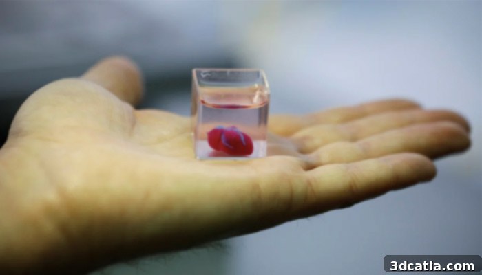
While the current 3D printed heart is modest in size, comparable to that of a rabbit’s heart, the underlying technology holds immense promise for scaling up. Prof. Dvir confirmed that the fundamental principles and techniques developed for this smaller prototype should be applicable to printing a full-sized human heart. The challenge, however, lies in ensuring the viability and functionality of larger, more complex structures. A key hurdle in tissue engineering is maintaining cell viability and integrating a functional vascular system that can supply nutrients and oxygen to every cell within the organ, while also removing metabolic waste. The achievement of creating a vascularized structure in this initial phase is therefore incredibly significant, laying the groundwork for future advancements.
Biocompatibility is a critical factor for the success of any implant. As Prof. Dvir explained, “The biocompatibility of engineered materials is crucial to eliminating the risk of implant rejection, which jeopardizes the success of such treatments. Ideally, the biomaterial should possess the same biochemical, mechanical, and topographical properties of the patient’s own tissues.” This personalized approach ensures that the printed organ integrates seamlessly with the patient’s body, functioning as naturally as a native organ. Matching these properties is vital for the long-term health and success of the transplanted organ, as it influences everything from cell adhesion and proliferation to overall tissue mechanics and responsiveness to biological signals.
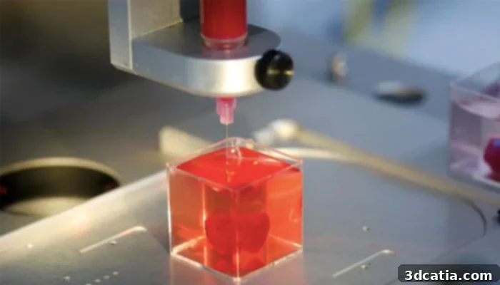
The researchers are now embarking on the crucial next phases of their study. The immediate goal involves culturing the printed hearts in the lab environment to further develop and “teach them to behave” like fully functional hearts. This incubation period will focus on maturing the cardiac muscle cells, allowing them to form stable connections and develop their electrical and mechanical properties. The aim is to achieve synchronized contractions that can effectively pump fluid, mimicking the natural action of a living heart. This process might involve electrical stimulation and mechanical conditioning to encourage the cells to organize into a coordinated, functional unit.
Following successful in-vitro development, the subsequent pivotal step will be to transplant these 3D-printed hearts into animal models. This phase will allow researchers to observe how the bio-printed organ behaves within a living system, assessing its functionality, integration, and long-term viability. “We need to develop the printed heart further. The cells need to form a pumping ability; they can currently contract, but we need them to work together,” concluded Prof. Dvir. This represents a significant challenge, moving beyond individual cell contractions to a cohesive, organ-level function. The insights gained from animal trials will be invaluable in refining the bioprinting techniques and ensuring the safety and efficacy of the technology before it can be considered for human clinical trials.
This pioneering research from Tel Aviv University is not merely about printing hearts; it represents a paradigm shift in regenerative medicine and personalized healthcare. The implications extend far beyond cardiac care, potentially paving the way for the creation of other complex organs, such as kidneys, livers, and lungs, which are also desperately needed for transplantation. The ability to custom-engineer organs that are genetically identical to the recipient could eliminate transplant waiting lists, eradicate the need for immunosuppressant drugs, and drastically improve the quality of life for millions of people worldwide. While challenges remain, including scaling up production, ensuring long-term organ function, and navigating regulatory pathways, this breakthrough serves as a powerful testament to human ingenuity and the boundless potential of 3D bioprinting technology to transform medicine as we know it.
This momentous achievement underscores the critical role of innovative scientific research in addressing some of humanity’s most pressing health issues. The journey from a rabbit-sized prototype to a fully functional human heart for transplantation will undoubtedly be long and complex, but the path has now been illuminated. It brings us closer to a future where life-saving organs can be printed on demand, custom-made for each patient, heralding a new era in personalized medicine and ensuring that heart disease, one day, may no longer be a death sentence.
What are your thoughts on this incredible new research and the future of 3D bioprinting? Let us know in a comment below or join the conversation on our Facebook and Twitter pages! And don’t forget to sign up for our free weekly Newsletter to get all the latest news in 3D printing delivered straight to your inbox!