Applications
STAMP and 3D Printing Used to Create Artificial Muscles for Medicine and Robotics
Artificial skeletal muscles are particularly important for the development of in vitro models for disease research or for use in biohybrid robots. However, there are some difficulties in the traditional production of these muscles, as 2D muscle monolayers often peel…
- 3 min read
- Applications
- Six more stories
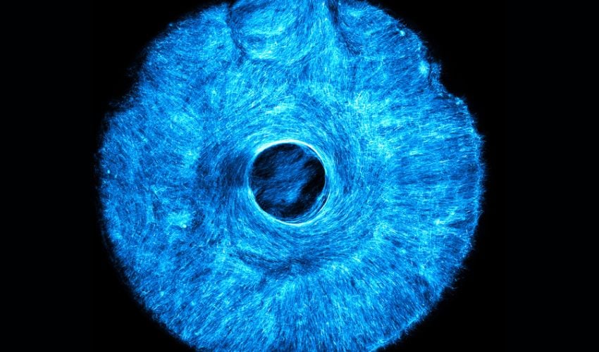
Artificial skeletal muscles are particularly important for the development of in vitro models for disease research or for use in biohybrid robots. However, there are some difficulties in the traditional production of these muscles, as 2D muscle monolayers often peel off after a few days and are not suitable for long-term use or live cell imaging – a technique in which cells are examined in a living state under a microscope or other imaging technique. For this reason, scientists at the Massachusetts Institute of Technology (MIT) have now applied a new technique to artificial muscle tissue that contracts in multiple directions and mimics movements of natural muscles. In the study, they presented a microtopographic stamping method that precisely controls the alignment of muscle fibers, and additive manufacturing played a particularly important role in this. The muscles could potentially be used in regenerative medicine, muscle disease research and robotics.
The new process “Simple Templating of Actuators via Micro-Topographical Patterning” (STAMP) uses 3D-printed stamps to structure microscopic grooves directly in natural hydrogels. The 3D-printed stamp is used to incorporate structured grooves into a soft hydrogel. These grooves guide the muscle cells as they grow and ensure that they align into functional fibers that can contract in different directions.
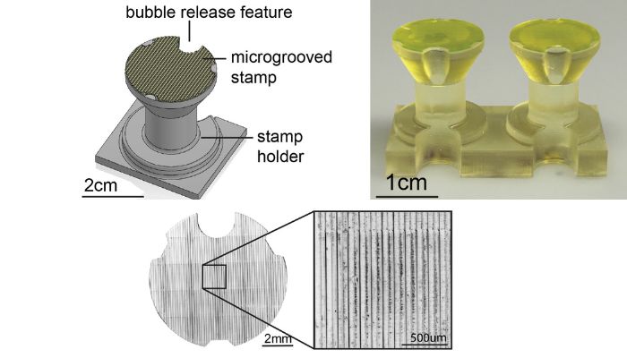
Top left: CAD model of the micro stamp and holder; top right and bottom: resulting 3D-printed parts.
But how exactly does the procedure work? The first step is to produce the 3D-printed stamps. These fit into 24-well plates and have fine vertical grooves (12.5-125 μm). The stamps are then fixed in a holder with bubble release and a liquid consisting of fibrinogen and thrombin is poured into the wells. The whole process is incubated at 37 °C for about an hour so that the fibrin polymerizes to form a cross-linked hydrogel. After removing the plunger, the hydrogel shows a precise, regular groove structure and a smoother surface, which improves the conditions for cell culture. The stamps are sterilized before use and can be reused several times after cleaning.
More from the desk
Six other notes from the rest of the bench.
 NewsRAYSHAPE Arrives in Europe, Expanding its 3D Printer Partners and ResellersAdditive manufacturing is an increasingly widespread technology worldwide and one of the continents experiencing considerable growth is Europe. The greatest European demand for 3D printing comes from small and medium-sized companies looking not only for prototyping, but also for
  NewsMatt Harkness is Recycling PLA Waste Using TattoosThe 3D printing filament PLA is often advertised as environmentally friendly since polylactic acid is largely made from renewable raw materials such as corn starch. Thus, products made from PLA are able to be labeled “biodegradable.” This eco-stamp can therefore&helli
 NewsP-Rouette, the 3D printed ballet slippersP-Rouette is the new invention of Hadar Neeman that should interest more than one ballet dancer. He has created 3D printed ballet slippers. This young graduate of the Academy of Art and Design of Bezalel has found the solution to most…
 News#3DExpress: Desktop Metal Announces Further Layoffs in New Cost Reduction Measures#3DExpress is our newest article format, which brings you some of the most interesting news from the week in a bite-sized format. This week, learn more about the new cost reduction plan announced by Desktop Metal, expected to start showing…
 PrintersReconstructing A 19th Century Ship in Australia Using 3D TechnologyWhile additive manufacturing offers the possibility of producing and repairing all sorts of parts, it can also be used to reconstruct old models, whatever they may be. And the Silentworld Foundation, an Australian non-profit organization focused on marine archaeology and…
 PrintersDa Vinci Color mini; the desktop colour 3D printerLast year, the manufacturer XYZprinting was talking about the colour additive manufacturing market with the arrival of its new 3D printer, the Da Vinci Color, a machine that combines InkJet technology and FDM, capable of printing up to 16 million shades…