
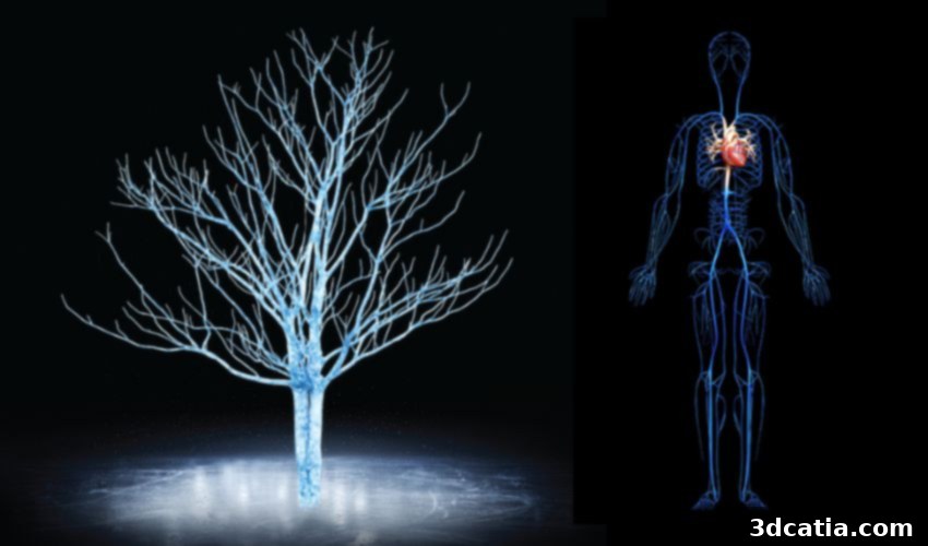
Bridging the Gap: Carnegie Mellon’s 3D Ice Printing Breakthrough for Artificial Organs and Functional Blood Vessels
The persistent and growing demand for organ transplants, particularly for vital organs like hearts, kidneys, and livers, presents one of the most significant challenges in modern medicine. For years, scientists worldwide have dedicated immense effort to developing viable artificial organs, yet tangible breakthroughs have been elusive, leaving a substantial gap between the number of available donor organs and the critical need for transplants. However, a revolutionary development from Carnegie Mellon University researchers offers a glimmer of hope. By pioneering a sophisticated 3D ice printing technique, these innovators have successfully engineered intricate structures that remarkably mimic natural blood vessels, promising a future where functional artificial organs could become a reality.
This timely announcement arrives amidst an escalating organ transplant crisis. According to the Division of Transplantation (DoT), the federal agency overseeing organ and blood stem cell transplant systems in the United States, the statistics are stark. As of 2024, a staggering 103,223 individuals—men, women, and children—were registered on the national transplant waiting list. This number is not static; it grows relentlessly, with a new person being added to the list approximately every eight minutes. This constant surge in demand, coupled with the limited supply of donor organs, underscores the urgent need for innovative solutions.
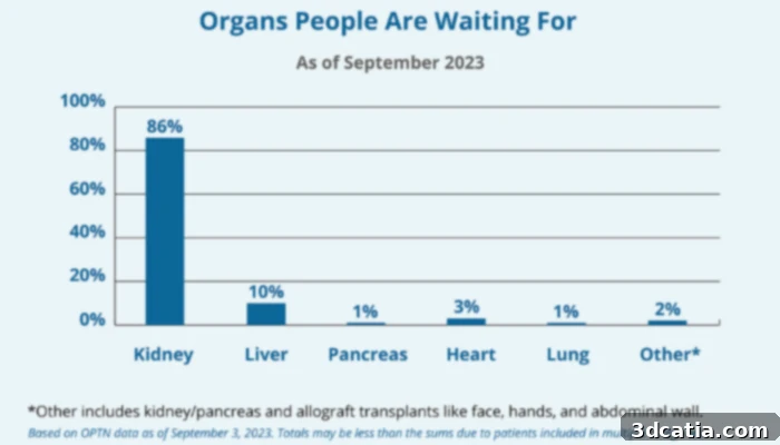
Many on the organ transplant list have waited for quite a while, especially for organs where demand is high, such as for kidneys (image credits: HRSA)
The human cost of this shortage is profound. The DoT estimates that approximately 17 people tragically die each day awaiting an organ transplant, translating to over 6,000 preventable deaths annually. This sobering reality fuels extensive research efforts, not only into increasing the number of organ donors—which stood at around 170 million registered individuals in the U.S. as of 2022, though actual donor viability varies—but also into exploring groundbreaking alternative methods to procure viable organs. Among these alternatives, 3D printing technologies have emerged as a beacon of hope, offering novel pathways to address this critical medical need.
While significant advancements have been made in bioprinting techniques over the years, aiming to create lab-grown organs, researchers have consistently encountered formidable obstacles. The most significant challenge lies in fabricating complex blood vessel networks within artificial organs that can replicate the intricate functionality of natural ones. Traditional approaches to designing artificial vascular systems often fall short, failing to mimic the precise architecture and dynamic responsiveness required for proper nutrient and oxygen exchange, as well as waste removal. This limitation has historically hindered the development of truly viable artificial organs. Now, with the advent of 3D ice printing, a transformative solution appears to be within reach.
3D Ice Printing: A Breakthrough for Artificial Blood Vessels and Organ Development
The innovative 3D ice printing technology, initially developed by engineering researchers at Carnegie Mellon University in 2022, represents a paradigm shift in bioprinting. The fundamental process involves precisely depositing a stream of water onto an extremely cold surface. What sets this technique apart is the use of “heavy water,” where the hydrogen atoms are replaced by deuterium. This modification not only elevates the water’s freezing point, allowing for greater control during the printing process, but also contributes to the creation of exceptionally smooth and uniform textures. As one might infer, the desired three-dimensional structure is meticulously built layer by layer through the controlled freezing of this specialized water.
Feimo Yang, a brilliant graduate student leading this groundbreaking research in the labs of Philip LeDuc and Burak Ozdoganlar at Carnegie Mellon University, offered deeper insights into what distinguishes 3D ice printing from other contemporary additive manufacturing processes. Yang explained, “What makes our method different from other kinds of 3D printing is that instead of letting the water completely freeze while we’re printing, we let it maintain a liquid phase on top. This continuous process, which is what we call freeform, helps us to get a very smooth structure. We don’t have a layering effect typical with many 3D printing techniques.” This critical difference – the ability to maintain a controlled liquid phase – is instrumental in achieving the remarkably smooth and continuous structures essential for replicating delicate biological networks, bypassing the inherent limitations of layered fabrication.
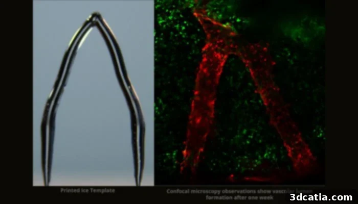
Image Credits: Feimo Yang
Once the intricate 3D printed ice templates are meticulously fabricated, they undergo a sophisticated post-processing step. These ice structures are carefully embedded within a biocompatible gelatin material known as GelMA. This composite is then exposed to ultraviolet (UV) light, a process that triggers the hardening of the gelatin matrix. Simultaneously, as the GelMA solidifies, the underlying ice template melts away, leaving behind perfectly formed, realistic blood vessel channels. This ingenious method ensures that the delicate internal architecture of the vasculature is preserved, resulting in channels that closely mirror the complexity and dimensions of natural capillary networks.
The efficacy of this pioneering approach has been demonstrated with significant success. The research team was able to successfully introduce endothelial cells – the specialized cells that line all blood vessels and play a crucial role in regulating exchanges between the bloodstream and surrounding tissues – into these newly fabricated blood vessels. Remarkably, these endothelial cells not only survived but thrived on the gelatin scaffolding for up to two weeks. This sustained viability is a monumental step forward, as it confirms the biocompatibility and functional potential of the engineered vascular networks. The researchers are now focused on culturing these cells for extended durations, aiming to observe their long-term functionality and integration within the artificial structures.
Beyond the monumental goal of creating fully functional artificial organs for transplant, this groundbreaking research holds immense promise for several other critical applications. The ability to precisely engineer complex, lifelike blood vessel networks represents a major advancement for tissue engineering, potentially enabling the creation of vascularized tissue patches for regenerative medicine. Furthermore, these sophisticated models could revolutionize pharmaceutical development by providing accurate *in vitro* platforms for testing the effects of drugs on blood vessels and vascular diseases. Such platforms could significantly reduce reliance on animal testing, accelerate drug discovery, and offer more precise insights into therapeutic efficacy and toxicity. This innovation from Carnegie Mellon University marks a truly transformative step forward, paving the way for unprecedented breakthroughs in the creation of artificial organs and advancing the frontiers of regenerative medicine and drug development.
This work not only offers a viable pathway to overcoming the chronic shortage of donor organs but also opens new avenues for studying vascular biology and pathology with unparalleled precision. The intricate control offered by 3D ice printing in creating microvascular networks could lead to a deeper understanding of various vascular diseases, including atherosclerosis, diabetes-related vascular complications, and even tumor angiogenesis. By recreating these conditions in a controlled laboratory setting, scientists can develop more targeted therapies and diagnostic tools. The potential implications for personalized medicine, where specific vascular models could be tailored to individual patients for disease modeling or drug screening, are also vast and exciting. You can learn more about this pivotal research in the official press release HERE.
What are your thoughts on the groundbreaking use of 3D ice printing to create artificial blood vessels? Do you believe this technology could be the crucial key to unlocking the future of artificial organ creation? We encourage you to share your insights and comments below, or engage with us on our LinkedIn, Facebook, and Twitter pages! Don’t miss out on the latest advancements in 3D printing – be sure to sign up for our free weekly newsletter here, delivering the freshest news directly to your inbox! Additionally, you can explore all our compelling videos on our YouTube channel for more in-depth content.
*Cover Image Credits: Carnegie Mellon University