
Revolutionizing Vision Research: How Bioprinting 3D Eye Tissues is Combating Macular Degeneration and Blinding Diseases
Vision, one of our five fundamental senses, is paramount to how humans and countless other species perceive and interact with the world. It allows us to navigate our environment, recognize loved ones, appreciate art, and gain knowledge. The sudden or gradual loss of sight can be profoundly debilitating, impacting an individual’s independence, quality of life, and overall well-being. Across the globe, millions suffer from visual impairment, with conditions ranging from cataracts and glaucoma to degenerative retinal diseases. In the United States alone, visual impairment affects an estimated 14 million people, posing a significant public health challenge. While some causes of vision loss are treatable, many, particularly degenerative eye diseases, remain incredibly difficult to correct, let alone cure. A major obstacle in understanding and developing treatments for these complex conditions is the limited availability of physiologically relevant human tissue samples for research and drug testing.
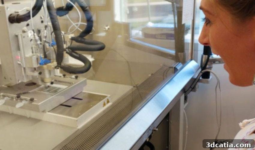
Addressing this critical need, researchers at the National Eye Institute (NEI), a vital component of the National Institutes of Health (NIH), which serves as the leading federal agency supporting medical research in the US, have pioneered an innovative approach: bioprinting functional eye tissues. This groundbreaking development aims to profoundly advance our understanding of blinding diseases, especially degenerative retinal conditions like age-related macular degeneration (AMD). While 3D printing technology has previously contributed to ophthalmology, such as the creation of a 3D printed eye prosthetic or the 3D printed cornea developed at the University of Newcastle, this NEI initiative marks a significant leap. It demonstrates the growing versatility of bioprinting for creating living, functional tissues specifically for disease modeling and therapeutic research, pushing the boundaries of what’s possible in the medical field.
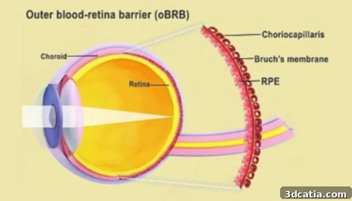
A diagram of the blood-retina barrier in the eye (photo credits: National Eye Institute)
Bioprinting’s Transformative Role in Advancing Research for AMD and Other Eye Diseases
Age-related macular degeneration (AMD) stands as a formidable challenge in eye health. It is a common and progressive condition predominantly affecting individuals aged 50 and older, and it represents the leading cause of vision loss among older adults globally. AMD targets the macula, the central part of the retina responsible for sharp, detailed central vision, crucial for tasks such as reading, driving, and recognizing faces. While AMD rarely leads to complete blindness, it causes a significant loss of central vision, manifesting as blurriness, distorted or wavy lines, and dark spots in the direct line of sight. This makes everyday activities incredibly difficult and frustrating for those affected, severely diminishing their quality of life. Nearly 20 million people in the US are estimated to have some form of AMD, and its prevalence continues to rise with an aging population.
There are two primary forms of AMD: dry (atrophic AMD) and wet (advanced neovascular AMD). Dry AMD, which accounts for about 85-90% of cases, progresses slowly and is characterized by the thinning of the macula and the presence of drusen (yellow deposits) under the retina. Wet AMD, though less common, is far more aggressive. It occurs when abnormal blood vessels grow under the retina, leaking fluid and blood, which leads to rapid and severe central vision loss. While various treatment options exist, especially for wet AMD, understanding the precise causes and the mechanisms that drive the progression from early stages to advanced dry and wet forms remains poorly understood. This lack of clear insight has long hampered the development of truly effective preventative measures and cures, highlighting the urgent need for better research models.
The NEI scientists, led by Kapil Bharti, Ph.D., who heads the NEI Section on Ocular and Stem Cell Translational Research, recognized these challenges and turned to bioprinting in conjunction with patient-derived stem cells. Their goal is to create more accurate and relevant human models that can significantly advance our understanding of the complex mechanisms underlying blinding diseases like AMD. Dr. Bharti and his team hope that this innovative bioprinting technique will provide a theoretically unlimited supply of patient-derived tissue. This is crucial because traditional research often relies on animal models, which, while useful, do not always accurately recapitulate the intricacies of human disease due to species-specific biological differences. The availability of patient-derived human tissue models offers an unprecedented opportunity to study degenerative retinal diseases in a highly relevant context, paving the way for more targeted and effective therapies.
Dr. Bharti explained the rationale behind their focus: “We know that AMD starts in the outer blood-retina barrier. However, mechanisms of AMD initiation and progression to advanced dry and wet stages remain poorly understood due to the lack of physiologically relevant human models. By printing cells, we’re facilitating the exchange of cellular cues that are necessary for normal outer blood-retina barrier anatomy. For example, presence of RPE cells induces gene expression changes in fibroblasts that contribute to the formation of Bruch’s membrane – something that was suggested many years ago but wasn’t proven until our model.” This highlights the profound impact of their work. The outer blood-retina barrier, comprising the retinal pigment epithelium (RPE) and Bruch’s membrane, plays a critical role in maintaining retinal health. Dysfunction in this barrier is a hallmark of AMD. By creating a functional model of this barrier, the researchers can observe cellular interactions and disease progression directly in a human-relevant system, confirming long-held hypotheses and opening new avenues for investigation.
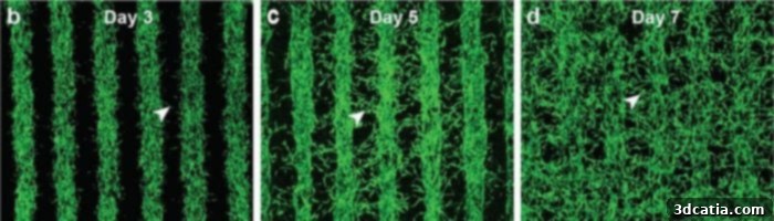
The progression of growth overtime for the cells (photo credits: Kapil Bharti)
To achieve these remarkable tissues, Dr. Bharti and his dedicated colleagues meticulously combined three immature choroidal cell types within a biocompatible hydrogel. The chosen cell types were pericytes, endothelial cells, and fibroblasts, each playing a crucial role in forming a functional tissue. Pericytes are vital cells that wrap around capillaries, regulating blood flow and maintaining the integrity of the blood-retina barrier. Endothelial cells are the building blocks of blood vessels, forming the inner lining of capillaries. Fibroblasts provide structural support and contribute to the extracellular matrix, which is the scaffolding that holds tissues together. This meticulously prepared cell-hydrogel mixture, often referred to as a “bio-ink,” was then precisely printed onto a biodegradable scaffold. This scaffold served as a temporary support structure, guiding the cells as they began to organize and mature, and is designed to degrade naturally over time, leaving behind the functional tissue.
The results were swift and promising. Within a matter of days following printing, the cells began to differentiate and mature, forming a dense capillary network, a critical component of the outer blood-retina barrier, as detailed in a press release about the project. The journey to full maturity for the bioprinted tissue took approximately 42 days. Upon reaching this stage, extensive tissue analyses, alongside genetic and functional testing, confirmed the viability and remarkable similarity of the printed tissues to native outer blood-retina barriers. This rigorous validation demonstrated that the bioprinted tissues not only looked anatomically similar but also behaved functionally like their natural counterparts. Even more critically for disease modeling, when subjected to induced stress, the printed tissue remarkably exhibited characteristic patterns indicative of early age-related macular degeneration. This capability to replicate disease pathology in a controlled environment makes these bioprinted tissues invaluable for studying AMD progression and testing potential therapeutic interventions.
Currently, the NEI researchers are actively utilizing these sophisticated bioprinted tissues to delve deeper into the complexities of AMD. This includes understanding the precise mechanisms of disease initiation, progression, and potential targets for intervention. Simultaneously, they are pushing the boundaries of the technology by experimenting with the addition of supplementary cell types to the printing process. This ongoing effort aims to create even more complex and accurate models that can recapitulate native eye tissue with greater fidelity, potentially incorporating photoreceptors or neural cells to build multi-layered retinal structures. This would further enhance the predictive power of these models for drug discovery and disease understanding.
Marc Ferrer, Ph.D., co-author of the study and director of the 3D Tissue Bioprinting Laboratory at NIH’s National Center for Advancing Translational Sciences, underscored the significance of this collaborative effort: “Our collaborative efforts have resulted in very relevant retina tissue models of degenerative eye diseases. Such tissue models have many potential uses in translational applications, including therapeutics development.” The implications for translational medicine are vast, ranging from high-throughput drug screening and personalized medicine approaches—where drugs can be tested on a patient’s own bioprinted tissue—to gaining unprecedented insights into genetic predispositions and environmental factors contributing to AMD. This breakthrough promises to accelerate the discovery of new treatments and ultimately bring hope to millions facing vision loss from degenerative eye conditions. You can find out more in the press release HERE or in the comprehensive scientific study published in Nature Methods HERE.
The development of bioprinted eye tissues by the NEI represents a monumental stride in ocular research, offering a powerful tool to unravel the mysteries of blinding diseases and accelerate the path to effective treatments. What are your thoughts on the revolutionary use of bioprinting to create functional eye tissues for research and potentially for future therapeutic applications? Share your insights and comments below, or engage with us on our LinkedIn, Facebook, and Twitter pages! Don’t forget to sign up for our free weekly Newsletter here to receive the latest 3D printing news directly to your inbox! You can also find all our videos and interviews on our YouTube channel for more in-depth content.