
Revolutionizing Pediatric Cardiology: The Promise of 3D Printed Biodegradable Stents
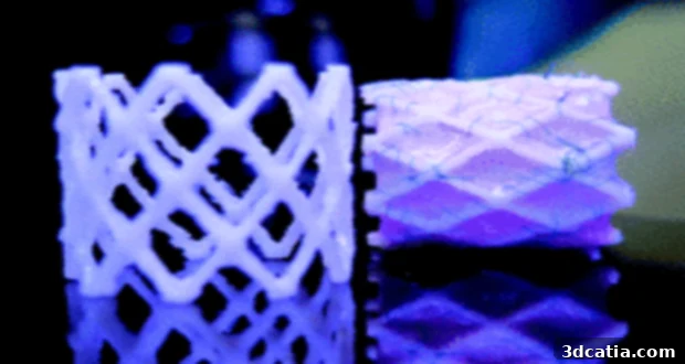
In a monumental stride towards advanced pediatric medical care, researchers at Eindhoven University of Technology (Tu/e) in the Netherlands have unveiled a groundbreaking innovation poised to transform the treatment of cardiovascular conditions in children. Leveraging the precision and versatility of 3D printing technology, this team has successfully developed a pioneering biodegradable stent, specifically engineered to address the unique challenges of growing pediatric patients. This medical device represents a significant leap forward from traditional stents, offering a future where interventions are less invasive and more harmonized with a child’s natural development.
The Critical Need for Pediatric Stents and Their Current Limitations
Stents are miniature mesh tubes, typically made from metal or plastic, that play a vital role in modern cardiology. Their primary function is to restore or maintain the patency of narrow or weakened arteries, often caused by the accumulation of plaque from conditions like atherosclerosis or congenital heart defects. By supporting the vessel walls, stents ensure adequate blood flow and can prevent life-threatening blockages. While highly effective in adult medicine, the application of conventional stents in pediatric patients presents a complex array of challenges that have long troubled medical professionals.
Current stents, whether fabricated from various metals like stainless steel and cobalt-chromium alloys or from non-degradable plastics, are designed for permanent implantation. This permanence becomes a significant drawback for children, whose bodies are constantly growing and developing. A stent implanted in an infant or young child will not grow with them, invariably leading to the need for multiple, invasive surgical procedures to replace or expand the device as the child ages. Each subsequent surgery carries its own risks, trauma, and recovery period, significantly impacting a child’s quality of life and placing an immense burden on families and healthcare systems.
Beyond the issue of growth, traditional stents are also associated with various clinical complications. One of the most prevalent is hyperplasia, an abnormal proliferation of tissue within the stented segment of the artery. This overgrowth can lead to restenosis, where the artery narrows again, negating the initial benefit of the stent. Other complications include chronic inflammation, potential for blood clot formation (thrombosis), and in some cases, the need for long-term anti-coagulant therapy, which carries its own risks. The metal or plastic materials, while biocompatible, are foreign bodies that remain in the circulatory system indefinitely, regardless of whether their structural support is still required.
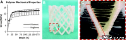
A: Stress–strain curves of polymer wire and dog bone samples; B: 3D printed prototype; and C: computational geometry adjusted considering the prototype dimensions. // Eindhoven University of Technology
Eindhoven University of Technology’s Groundbreaking Innovation
Understanding these critical limitations, researchers at Tu/e embarked on a mission to redefine pediatric cardiovascular intervention. Their solution: a novel 3D printed biodegradable stent. This ingenious device is engineered not to remain permanently, but to gradually and safely dissolve within the body over time, eliminating the need for subsequent removal surgeries. This design philosophy is particularly revolutionary for children, as it allows their developing vascular systems to heal and grow unimpeded, free from the constraints of a non-degradable implant.
The concept of a biodegradable stent offers a paradigm shift in treatment. Instead of a lifelong foreign object, the stent serves its temporary purpose of supporting the vessel during healing, and once the artery has recovered sufficient strength, the stent naturally breaks down into harmless compounds that are safely absorbed by the body. This adaptive approach not only minimizes long-term complications but also significantly reduces the physical and psychological burden on pediatric patients and their families, promising a future of fewer medical procedures and a better quality of life.
The Science Behind the Solution: 3D Printing and Advanced Materials
The success of the Tu/e team hinges on the strategic integration of advanced manufacturing techniques with cutting-edge biomaterials. Additive manufacturing, commonly known as 3D printing, proved to be the ideal technology for creating these intricate and customizable medical devices. 3D printing allows for the fabrication of complex geometries with high precision, which is crucial for stents that must conform perfectly to individual arterial anatomies. This level of customization is difficult, if not impossible, to achieve with traditional manufacturing methods for such small-scale, delicate structures.
Precision Manufacturing with Additive Technology
For the actual fabrication of their prototypes, the researchers employed a MakerBot Replicator 2x. This choice highlights the increasing accessibility of advanced manufacturing tools for research and development. Using such a desktop FDM (Fused Deposition Modeling) printer enabled rapid prototyping and iteration, allowing the team to quickly test and refine their designs. The ability to produce stents on demand with specific dimensions tailored to a child’s unique vascular structure is a significant advantage, potentially paving the way for truly personalized medicine in pediatric cardiology.
Strategic Material Selection for Optimal Integration
The material selection for the stent was paramount to its success. Initially, a model of the stent was created using nitinol, a shape-memory alloy known for its superelasticity and biocompatibility, often used in medical devices where flexibility and resilience are critical. This nitinol model likely served as a design template or a benchmark for mechanical properties. The actual prototype stents, however, were developed using a flexible copolyester elastomer. The incorporation of this specific material was not accidental; its properties were carefully chosen to promote the long-term acceptance of the medical device within the body. Copolyester elastomers offer excellent biocompatibility, meaning they are well-tolerated by biological systems and do not elicit adverse immune responses. Their inherent flexibility allows the stent to adapt to the dynamic environment of a blood vessel, while their controlled biodegradability ensures that the stent gradually disappears once its structural support is no longer needed.
This careful selection of a flexible, biodegradable material directly addresses the core problem of pediatric stents: the need for a device that accommodates growth. By slowly degrading, the stent gently allows the natural artery to remodel and expand as the child grows, avoiding the need for repeated surgical interventions that are currently mandatory with permanent implants. This self-resolving nature means the child’s body can gradually take over the supportive function, ultimately leaving behind no permanent foreign material.
Rigorous Testing for Safety and Efficacy
To validate the functionality, stability, and biodegradability of their innovative stents, the Tu/e researchers subjected their prototypes to a comprehensive battery of tests. These experiments were crucial for understanding how the stent would perform under physiological conditions and how its material would behave over time within the body. The testing phase is a critical step in any medical device development, ensuring both safety and efficacy before potential clinical applications.
Among the tests conducted were computational crush and crimping tests. Computational modeling allowed the researchers to simulate the mechanical stresses and deformations the stent would experience during implantation and once in place within an artery. Crush tests evaluate the stent’s radial strength—its ability to resist external compression and maintain the vessel lumen. Crimping tests are equally vital, assessing how effectively the stent can be compressed onto a delivery catheter and then expanded to its intended diameter within the artery. This process requires significant mechanical integrity, as the stent must withstand the forces of delivery through often tortuous and narrow vessels, then deploy reliably to hold the artery open. These tests are paramount to ensuring the stent can be delivered safely and effectively without permanent deformation or damage.
Crucially, the team also performed extensive degradation tests to ascertain the stent’s biodegradability profile. Utilizing accelerated hydrolysis, they simulated the degradation process that would occur in the human body over an extended period. This method allows researchers to predict the rate at which the stent material breaks down and to ensure that the degradation products are non-toxic and safely absorbed by the body. Controlling the degradation rate is vital: the stent must maintain its structural integrity long enough for the artery to heal and strengthen, but then degrade completely before it becomes an obstruction or source of chronic inflammation. These tests provide invaluable data on the stent’s lifespan and its interaction with biological fluids.
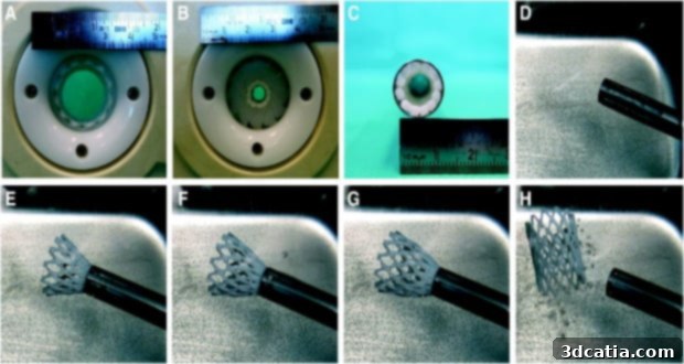
Crimping and delivery of the 3D printed prototype. A: 3D printed stent placed inside crimping device. B: Stent crimping to an approximate diameter of 10 mm. C: Stent transferred to the transapical delivery device with an internal diameter of 12 mm. (D–H) Self-expansion as the stent is pushed out of the delivery system. // Eindhoven University of Technology
The Road Ahead: Future Developments and Potential Impact
While the initial results are exceptionally promising, the 3D printed biodegradable stents from Tu/e are still in the concept stage. This means there is further research and development required before these devices can be translated into clinical practice. However, this new innovation unequivocally offers a powerful and viable alternative to the current medical devices used for pediatric cardiovascular interventions. The extensive tests and demonstrations conducted on these prototypes lay a robust foundation for future advancements.
The researchers are now focused on refining the technology, primarily tasked with two key areas: identifying and integrating even better materials, and reducing imperfections in the struts’ profile. Finding superior biomaterials involves exploring polymers with tailored degradation rates, enhanced mechanical strength, and even bio-active properties that could promote healing or reduce the risk of restenosis. The goal is to optimize the material composition to perfectly match the healing timeline of various cardiovascular conditions in children, ensuring optimal support followed by complete and harmless dissolution. Reducing imperfections in the strut profile, the individual structural elements of the stent, is equally crucial. Smoother struts can minimize turbulence in blood flow, thereby reducing the risk of blood clot formation and further limiting the potential for tissue irritation or hyperplasia. Achieving micron-level precision through advanced 3D printing techniques will be central to this refinement.
The potential impact of this research extends far beyond pediatric cardiology. The principles of 3D printing custom, biodegradable implants could revolutionize treatment across various medical disciplines. Imagine personalized implants for bone regeneration, nerve guides, or even tissue scaffolds that naturally integrate and disappear as the body heals. For pediatric patients with cardiovascular issues, this technology promises a future where a single intervention could suffice, eliminating the need for traumatic, repeated surgeries throughout their childhood and adolescence. This not only improves their physical health but also their psychological well-being, allowing them to lead more normal, active lives. The journey from concept to clinic will involve rigorous regulatory approvals and extensive clinical trials, but the vision of a truly adaptive and growth-friendly stent is now closer than ever before, thanks to the pioneering work at Eindhoven University of Technology.
You can read the full report and delve deeper into the scientific details of this remarkable innovation here.
What are your thoughts on these cutting-edge 3D printed biodegradable stents? Do you believe this medical device holds the key to significant advancements in pediatric cardiology and beyond? Share your insights and perspectives in a comment below or join the conversation on our Facebook and Twitter pages!