
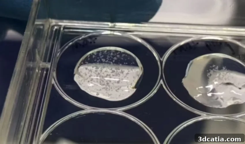
Revolutionizing Pregnancy Research: The Power of Bioprinted Placental Organoids
The placenta, often described as a temporary organ, is in fact a remarkably sophisticated and indispensable component of human gestation. Possessing a multitude of unique traits, it is the sole human organ that is “disposable,” forming anew with each successful implantation of a fertilized egg within the uterus. Following conception, it embarks on a journey of rapid growth, developing in perfect synchronicity with the fetus, eventually reaching the substantial size of a small plate. This extraordinary organ serves as the baby’s literal lifeline, fulfilling crucial roles that are vital for survival in the womb. Since babies do not breathe or consume food directly, the placenta acts as their primary source of oxygen and essential nutrients. Beyond this, it performs the critical functions of the baby’s kidneys and lungs, meticulously filtering metabolic waste products from the fetal bloodstream and facilitating the exchange of gases. Furthermore, the placenta plays a pivotal role in producing hormones necessary to sustain pregnancy and acts as an immune barrier, protecting the fetus from potential pathogens while allowing beneficial antibodies to pass through.
Despite its indisputable criticality to successful fetal development and maternal health, the human placenta remains one of the least understood and most enigmatic organs in human biology. This significant gap in knowledge stems from several inherent challenges in studying it. One primary difficulty lies in the ethical and practical complexities of obtaining samples during pregnancy. Invasive procedures to acquire placental tissue carry inherent risks, including potential complications such as infection, hemorrhage, or, tragically, miscarriage. These risks severely limit the frequency and scope of research during the most crucial developmental stages. Once a baby is born, the placental tissue undergoes rapid and significant structural and functional changes, rendering it unsuitable for studying the early and dynamic processes of placental development and function. Moreover, while animal models are invaluable in many areas of biomedical research, they provide only limited insights into the human placenta. The placentas of most animal species differ substantially in structure, cellular composition, and physiological function from that of humans, making direct extrapolation of findings problematic. Consequently, researchers continue to grapple with a fundamental lack of comprehensive understanding regarding how the human placenta develops, functions, and critically, how it malfunctions, particularly during the intricate early stages of pregnancy when many complications originate.
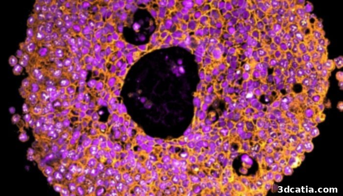
A placental organoid under the microscope
Placental Organoids: A New Frontier in Research
In an innovative effort to bridge this critical knowledge gap, researchers, notably those from the University of Technology Sydney, are leveraging cutting-edge technology to create artificial mini-placentas, known as organoids. These sophisticated 3D cellular structures are designed to mimic the complex architecture and functionality of real organs on a microscopic scale. The concept of organoids first emerged on the medical research scene around 2009, quickly gaining recognition for their potential to revolutionize disease modeling and drug discovery. Typically, organoids are generated by carefully suspending pluripotent stem cells or adult stem cells in a specialized gel or extracellular matrix. This unique environment allows the cells to self-organize, grow, and divide, forming three-dimensional clusters that recapitulate many aspects of an actual organ’s structure and function. In a significant milestone for placental research, the first placental organoids were successfully created in 2018, utilizing trophoblasts – a unique and specialized cell type found exclusively within the placenta, essential for its development and function.
The ability to cultivate and study placental organoids in a controlled laboratory setting offers an unprecedented opportunity to delve into crucial processes of early pregnancy. This innovative approach holds immense promise for uncovering the underlying causes of serious and often devastating conditions, such as preeclampsia. Preeclampsia, a severe pregnancy complication characterized by high blood pressure and signs of damage to other organ systems, most often the liver and kidneys, is intrinsically linked to placental dysfunction. Affecting a significant 5-8% of all pregnancies globally, preeclampsia represents a major public health concern. The condition is responsible for a staggering 46,000 maternal deaths and approximately 500,000 newborn deaths worldwide each year, underscoring its devastating impact. Even when not fatal, preeclampsia can lead to severe high blood pressure, protein in the urine, and can cause irreparable damage to vital maternal organs, including the kidneys, liver, brain, and blood system. Furthermore, it frequently necessitates early delivery, posing significant risks and long-term health challenges for both mothers and babies, including increased risk of cardiovascular disease for the mother and developmental issues for the child. Tragically, the only definitive “cure” for preeclampsia currently available is the delivery of the baby and placenta, precisely because scientists still lack a comprehensive understanding of its precise causes and mechanisms, a void that placental organoids aim to fill.
While the advent of placental organoids marked a significant leap forward, previous efforts to study the placenta through these models faced their own set of limitations. Most of these early studies relied on suspending trophoblasts in gels derived from animal sources, such as Matrigel. While functional, these animal-derived gels present several drawbacks. Critically, they do not accurately or consistently reflect the complex and dynamic cellular microenvironment of an actual human placenta. Their batch-to-batch variability can introduce inconsistencies in experimental results, and their undefined composition makes it challenging to pinpoint specific factors influencing organoid development. Moreover, the manual process of suspending cells to create a sufficient quantity of organoids for robust research is often cumbersome, labor-intensive, and difficult to scale, thereby limiting the throughput and scope of experiments.
Why Bioprinting is the Future of Placental Research
The placental organoids developed by the pioneering team at the University of Technology Sydney represent a monumental advancement because they are the first of their kind to be successfully bioprinted. This distinction is crucial, as bioprinting offers a level of precision and control simply unattainable with traditional organoid culture methods. Bioprinting utilizes advanced additive manufacturing techniques to precisely deposit cells and biomaterials layer by layer, creating intricate 3D tissue structures. Furthermore, a key innovation in the UTS study was the use of a novel mix of trophoblast cells combined with a synthetic, highly controllable gel, rather than the less reliable and variable animal-based gels. This synthetic hydrogel provides a more defined and reproducible environment, allowing researchers to meticulously control the physical and biochemical cues that influence cell behavior and tissue development.
The results of this groundbreaking bioprinting approach were immediately apparent and highly significant. These bioprinted organoids exhibited distinct growth patterns and developed a different, and crucially, more relevant number of cell sub-types compared to those grown in traditional animal-derived gels. This indicates that the bioprinting process, coupled with the synthetic matrix, creates an environment that better mimics the native human placenta. Claire Richards and Lana McClements, who were co-authors of the seminal study, highlighted the profound implications of their research in an article for The Conversation. They eloquently stated, “These organoids were very similar to human placental tissue, providing an accurate model of the early placenta.” This remarkable fidelity to human tissue is a game-changer for research. They further elaborated on the dynamic nature of their model, noting, “We could change how the cells organised themselves by taking young organoids out of the gel and letting them float in their liquid food.” This ability to manipulate the cellular architecture and observe subsequent changes in real-time opens up unparalleled avenues for understanding fundamental placental development and potential dysfunctions, offering a dynamic window into this previously elusive organ.
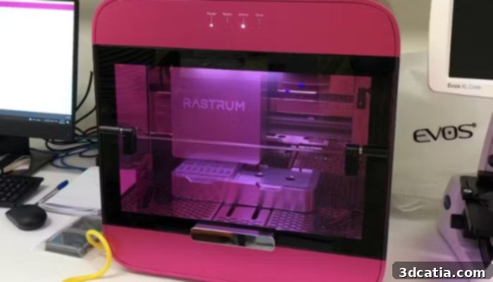
Bioprinters are advantageous because they allow cells to be positioned precisely in a 3D structure
Transformative Potential Applications
The development of highly accurate placental organoids, particularly those generated through advanced bioprinting techniques, holds immense potential to revolutionize our understanding and management of pregnancy complications. These miniature organs provide an unprecedented platform for researchers to deeply investigate the intricate mechanisms behind conditions like preeclampsia, gestational diabetes, and intrauterine growth restriction. For instance, the pioneering Sydney researchers conducted a compelling experiment where they exposed their bioprinted organoids to a specific immune signal known to be highly present in women diagnosed with preeclampsia. By meticulously observing the organoids’ responses and then testing various therapeutic treatments, they demonstrated the direct applicability of these models for screening potential interventions and gaining crucial insights into disease progression. This ability to mimic disease states in a controlled environment is invaluable for preclinical research.
Looking ahead, the integration of powerful genomic editing tools such as CRISPR technology with placental organoids could unlock even deeper levels of understanding. Researchers could precisely edit specific genes within the organoid cells to investigate their roles in placental development and disease, thereby unraveling the genetic underpinnings of various pregnancy complications. Beyond genetic studies, these sophisticated organoids can be utilized to examine the effects of infections, such as Zika or rubella, on placental function and fetal health, providing a safer and more ethical alternative to animal models or human studies. Furthermore, placental organoids are perfectly poised to serve as robust platforms for rigorously testing the safety and effectiveness of new drugs and therapies intended for pregnant women. This is particularly critical given the historical exclusion of pregnant individuals from many clinical trials, leaving a significant gap in our knowledge of drug efficacy and safety during pregnancy. With bioprinting, scientists can significantly improve the accuracy, reproducibility, and scalability of their experiments, accelerating the pace of discovery and reducing the ethical concerns and costs associated with animal testing.
Ultimately, this cutting-edge technology is set to play a pivotal and transformative role in improving maternal and fetal health outcomes globally. By providing accurate, controllable, and scalable models of the human placenta, bioprinted organoids empower researchers to predict, prevent, and treat pregnancy complications with unprecedented precision. This means a future where early detection of risks is more reliable, where targeted therapies can be developed and validated faster, and where the health of both mothers and their babies is safeguarded more effectively than ever before. To delve deeper into this fascinating research and its implications, we encourage you to read the full article HERE.
What are your thoughts on the incredible potential of 3D printed placentas in advancing medical science? We’d love to hear your perspective! Please share your insights in a comment below or connect with us on our LinkedIn or Facebook pages! Don’t forget to sign up for our free weekly Newsletter to receive the latest updates and breaking news in the world of 3D printing directly to your inbox. You can also explore all our informative videos and engaging content on our YouTube channel. Interested in more news and developments in medical and dental 3D printing? Visit our dedicated page HERE for a comprehensive overview of the latest innovations in this rapidly evolving field.
*Cover Image: Organoids suspended in gel, ready to slice and view under the microscope. All Photo Credits: Claire Richards