
Revolutionizing Orthopedic Surgery: The Breakthrough 3D-Printed Femur Model for Enhanced Surgical Planning and Research
The rapid advancements in 3D printing technologies are profoundly transforming various sectors, with its application in medicine experiencing an unprecedented boom. Among the most exciting innovations, researchers at the University of Texas at Dallas (UT Dallas), in a remarkable collaboration with orthopedic surgeons at UT Southwestern Medical Center, have successfully designed and developed a highly realistic 3D-printed femur model. This groundbreaking project is poised to provide medical professionals with an invaluable new tool, fundamentally changing how doctors prepare for intricate bone reconstruction surgeries and develop more effective treatments for challenging conditions like bone tumors. The initial findings and methodology of this significant study have been officially published in the esteemed “Journal of Orthopaedic Research,” marking a pivotal moment in the convergence of additive manufacturing and medical science.
The primary goal behind the creation of this sophisticated 3D-printed femur is to facilitate and enhance bone reconstruction surgery, offering a tangible, patient-specific replica for pre-operative planning. The research specifically focused on the central shaft of the femur, meticulously defining the precise parameters required for producing a 3D model that is robust and accurate enough for rigorous biomechanical testing. This innovative approach allows surgeons to practice complex procedures, identify optimal implant placements, and anticipate potential challenges before entering the operating room, thereby significantly reducing surgical risks and improving patient outcomes. While the potential benefits and implications of this technology are undeniably promising, it is crucial to recognize that it represents an early stage of innovation. Before it can be widely adopted in clinical settings and integrated into routine surgical practice, several additional stages of comprehensive research, validation, and stringent regulatory approval will be necessary to ensure its safety, efficacy, and widespread applicability across diverse patient populations.
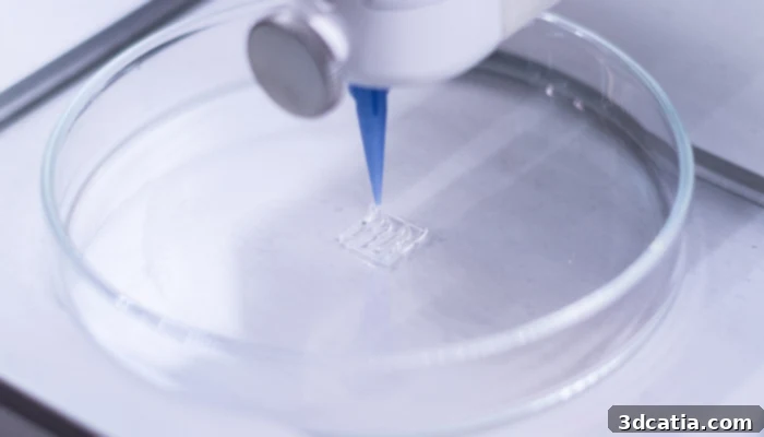
Detailed femur samples produced using advanced 3D printing technology for orthopedic study.
Transforming Biomechanical Studies with Patient-Specific 3D Printing
Traditionally, orthopedic surgeons and researchers have relied on conventional methods to examine and thoroughly evaluate new surgical techniques, implant designs, and fixation strategies. These methods primarily involve the use of human cadaveric donor bones or commercially available synthetic bone models. While these approaches have served as foundational tools, enabling surgeons to identify optimal fixation methods and anticipate bone reactions, they come with a significant set of inherent limitations. The acquisition and preparation of donor bones are often incredibly costly and time-consuming, involving ethical considerations, storage challenges, and inherent biological variability that can affect study consistency. Moreover, both donor bones and generic synthetic models inherently lack the ability to be tailored to the unique anatomical complexities and specific pathological conditions of individual patients. This limitation significantly curtails their effectiveness in highly personalized clinical situations. This generic approach often means that solutions developed cannot fully account for the vast variability in human bone structure, density, and pathology, thereby posing challenges in achieving truly optimized and individualized outcomes for every patient requiring orthopedic intervention.
Addressing the Challenges with Innovative Additive Manufacturing
Recognizing these critical challenges, the forward-thinking researchers and clinicians at UT Southwestern proactively sought a more innovative, efficient, and cost-effective solution for conducting advanced biomechanical studies and pre-surgical planning. This imperative led them to establish a crucial collaboration with Dr. Wei Li, a distinguished 3D printing specialist based at UT Dallas, renowned for his expertise in additive manufacturing. Dr. Li’s profound knowledge and practical experience in 3D printing proved to be the missing link in developing a rapid, economical, and highly customizable alternative to traditional, restrictive methods. Dr. Wei Li succinctly articulates the core need from a surgeon’s perspective: “To make plans for surgery, surgeons absolutely need to know the exact and intricate geometry of the bone they are working on.” He elaborates on the revolutionary potential of their collaborative work, stating, “With advanced 3D printing technology, we’re now able to print out the femur bone sample with the same intricate geometry as the actual femur inside a patient’s body. This unprecedented level of precision and patient-specificity opens up entirely new avenues for pre-surgical planning, advanced biomechanical research, and personalized medical education.” This ability to replicate exact anatomical structures from patient imaging data offers an unparalleled advantage, facilitating patient-specific planning that was previously unimaginable with generic models.
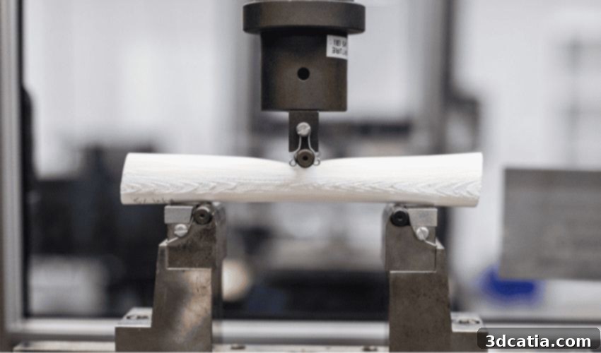
The practical development and meticulous refinement of these sophisticated 3D-printed models were expertly spearheaded by Kishore Mysore Nagaraja, a dedicated PhD student in mechanical engineering at UT Dallas. Working diligently under the direct supervision and invaluable guidance of Dr. Wei Li in his advanced manufacturing laboratory, Nagaraja embarked on an intensive, iterative process of trial and error. This methodical approach involved meticulously designing, printing, and refining several successive versions of the 3D-printed femur model. Each prototype underwent rigorous and comprehensive mechanical performance tests, specifically designed to assess its durability, flexibility, fracture resistance, and overall structural integrity under simulated physiological stresses. The overarching objective of these exhaustive and precise tests was to ensure that the artificial bone models would perform as closely as possible to natural human femurs in terms of their biomechanical response. This meticulous approach guaranteed that the models were not just visually accurate representations but also functionally representative of human bone, a critical factor for their utility in reliable biomechanical research, effective surgical rehearsal, and ultimately, improved patient outcomes.
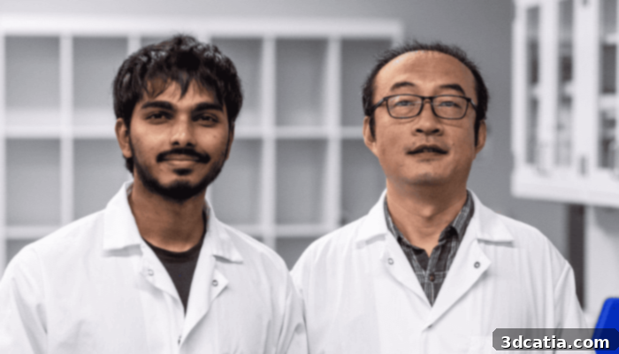
Mechanical engineering PhD student Kishore Mysore Nagaraja (left) and Dr. Wei Li (right) demonstrating their innovative 3D-printed femur model.
Affordable and Accurate: The Power of Polylactic Acid (PLA) in Medical Modeling
For the creation of these highly realistic and functional bone replicas, the researchers strategically opted for polylactic acid (PLA), a versatile, affordable, and notably environmentally friendly polymer. PLA is a thermoplastic polyester derived from renewable resources like corn starch or sugarcane, making it a sustainable choice compared to petroleum-based plastics. Its widespread use in consumer-grade 3D printing, particularly FDM (Fused Deposition Modeling) technology, is primarily due to its ease of processing, good mechanical properties, and relatively low material cost. In this medical application, PLA’s specific characteristics proved ideally suited for the task; it allowed for the rapid and economical production of models while possessing sufficient mechanical strength, rigidity, and biocompatibility to mimic human bone effectively for testing purposes. The resulting model, faithfully representing the central diaphysis (shaft) of the human femur, measures approximately 20 centimeters in length and 2.5 centimeters in diameter, carefully scaled to accurately reflect the anatomical proportions relevant for detailed biomechanical assessment and surgical planning.
The meticulous biomechanical tests conducted on these PLA replicas yielded truly remarkable and validating results. The 3D-printed models consistently demonstrated mechanical performance comparable to that of actual human femurs, a critical validation of their immense potential utility in both surgical planning and advanced orthopedic research. This means the models can accurately withstand similar forces, exhibit analogous deformation characteristics, and respond to stress in a manner consistent with real bone, thereby providing reliable and actionable data for evaluating various surgical approaches, implant efficacy, and patient-specific biomechanics. Beyond its impressive structural and functional performance, one of the most compelling aspects of this innovation is its astonishing cost-effectiveness. The production cost of a single 3D-printed femur model is estimated to be around a mere $7. This drastically low cost stands in stark contrast to the significantly higher expenses associated with acquiring and preparing cadaveric specimens or purchasing proprietary synthetic bones, making advanced surgical planning, personalized research, and comprehensive medical education far more accessible to a wider range of medical institutions, hospitals, and educational programs globally. This affordability could democratize access to high-fidelity anatomical models, fostering innovation and training across the medical field.
Expanding Horizons: Diverse Applications and Future Potential
Dr. Wei Li enthusiastically emphasizes the broad spectrum of transformative applications for these versatile 3D-printed bone models, extending far beyond just basic pre-surgical planning. For instance, the research opens up exciting possibilities for the polymer used, or similar advanced biodegradable and biocompatible materials, to eventually replace traditional, inert materials such as titanium in certain bone repair procedures. Imagine implants that can gradually degrade and resorb into the body as the natural bone heals and regrows, or intricate scaffolds that actively encourage host tissue integration without the need for secondary removal surgeries. Furthermore, Dr. Li highlights the innovative potential of printing patient-specific tumors directly onto these highly accurate bone models. This would create highly realistic, personalized replicas of cancerous bone structures, allowing oncological surgeons and researchers to meticulously evaluate different treatment regimens, plan complex tumor resections with unprecedented precision, and even test novel drug delivery systems or radiation therapies in a precise, risk-free, and patient-specific environment before applying them to actual patients. This could lead to significantly more effective, less invasive, and highly targeted cancer treatments, ultimately improving prognoses and quality of life for cancer patients.
Another incredibly exciting avenue of exploration involves utilizing these precisely engineered replicas to actively promote the regeneration of human bone tissue. These 3D-printed scaffolds, tailored to individual patient needs, could serve as biomimetic templates, providing the ideal microenvironment to guide the growth of new bone cells and facilitating the natural healing process in severe defects or non-union injuries. The ability to create custom-shaped, porous structures with specific mechanical properties and integrated biological cues could revolutionize regenerative medicine, particularly for patients suffering from significant bone loss due to trauma, disease, or congenital conditions. The profound impact of this research also extends significantly to medical education and training. These affordable, accurate, and patient-specific models can become indispensable teaching tools for medical students, residents, and even seasoned surgeons, allowing them to gain invaluable hands-on experience with complex anatomical structures and intricate surgical procedures in a simulated, consequence-free environment. This practical, immersive training can dramatically improve surgical proficiency, reduce learning curves for new techniques, and ultimately translate to safer and more effective care for future patients.
As this cutting-edge technology matures and gains wider acceptance, its seamless integration into various facets of medical practice could lead to the realization of truly personalized medicine on an unprecedented scale. Surgeons could perform “dry runs” of complex, high-stakes surgeries on an exact replica of a patient’s unique bone structure, anticipating potential challenges, identifying anatomical anomalies, and meticulously refining their surgical approach. This pre-operative rehearsal can lead to significantly reduced operating times, fewer intra-operative complications, and substantially improved long-term patient outcomes. Moreover, the detailed feedback garnered from rigorous biomechanical testing on these highly accurate models can directly inform the design and optimization of next-generation implants, prosthetics, and advanced surgical tools, continually pushing the boundaries of what is possible in orthopedic care and medical innovation. The exemplary collaboration between the engineering expertise of UT Dallas and the clinical insights of UT Southwestern Medical Center underscores the immense power of interdisciplinary research in driving medical innovation forward at an accelerated pace. While there are still vital steps to be taken for widespread clinical translation and regulatory approval, the robust foundation laid by this project is immensely promising and holds tremendous potential for shaping the future of orthopedic, oncological surgery, and personalized healthcare for decades to come.
We invite you to share your valuable thoughts and insights on this remarkable 3D-printed femur innovation! How do you foresee this cutting-edge technology impacting the future of orthopedic surgery, patient care, and personalized medicine? Let us know in a comment below or join the vibrant conversation on our LinkedIn, Facebook, and Twitter pages! Don’t miss out on the latest advancements and breaking news in additive manufacturing – sign up for our free weekly newsletter here to receive cutting-edge 3D printing news directly to your inbox. You can also explore all our insightful videos and in-depth discussions on our dedicated YouTube channel, where innovation comes to life!
*All Photo Credits: The University of Texas at Dallas