
3D Printed Human Ear: A Breakthrough in Reconstructive Surgery
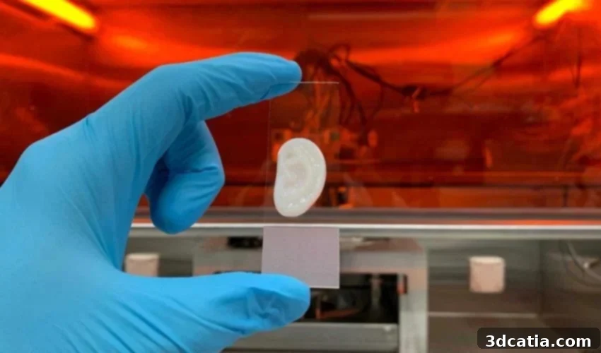
The field of regenerative medicine is constantly pushing the boundaries of what’s possible. While the vision of readily available 3D printed organs for transplant remains a long-term aspiration, significant strides are being made in laboratories worldwide. One notable advancement comes from a team of researchers at ETH Zurich, who have successfully engineered elastic, stable ear cartilage from human cells grown in the lab. This achievement, led by researcher Philipp Fisch and Professor Marcy Zenobi-Wong, brings personalized solutions for patients with severe ear malformations or injuries a crucial step closer to reality.
Addressing the Limitations of Current Ear Reconstruction Techniques
The primary motivation behind this research is to overcome the inherent limitations of existing ear reconstruction methods. Currently, the standard procedure for addressing ear malformations or ear loss due to accidents involves harvesting cartilage from the patient’s own ribs to meticulously sculpt a new ear. While this approach can be effective, it presents several drawbacks. Firstly, it is an invasive and painful procedure for the patient. Secondly, the resulting ear often lacks the natural flexibility and elasticity of a native ear, leading to a less than ideal outcome.
Scientists are therefore exploring alternative approaches that involve creating a tissue engineered ear that possesses both the necessary structural stability and the flexibility required for a natural appearance and feel. The ETH Zurich team’s work represents a significant advancement in this direction.
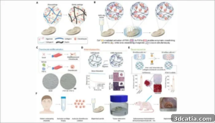
Schematic overview of the ear cartilage manufacturing process (credits: P. Fisch, S. Kessler, S. Ponta, et al.).
The Process: From Cell Extraction to 3D Printing
The creation of the 3D printed ear cartilage involves a sophisticated multi-step process, starting with obtaining the necessary biological material. Researchers extracted cells from small cartilage fragments that were leftover from previous surgeries performed on other patients. This approach minimizes the need for invasive procedures solely for research purposes. From a small initial sample measuring just three millimeters, the team isolated approximately 100,000 cells.
These extracted cells were then cultured and expanded in a specialized nutrient solution. The goal was to cultivate a sufficient quantity of cells – in the hundreds of millions – needed to construct a complete ear structure. During this crucial cultivation phase, the research team meticulously optimized the environment to encourage the cells to produce type II collagen and elastin, both essential components characteristic of ear cartilage. Preventing the cells from differentiating into fibroblasts, which would produce softer scar tissue, was a critical aspect of this process. The precise control over the cellular environment is vital for ensuring the desired tissue properties.
Bio-ink and 3D Printing the Ear Structure
Once the cell population had been sufficiently expanded and the cells were producing the desired extracellular matrix components, the cells were mixed with a specialized “bio-ink”. This bio-ink is essentially a gelatinous material that serves as a support matrix for the cells, providing them with a three-dimensional environment in which to organize and develop. This bio-ink is carefully formulated to be biocompatible and to provide the necessary mechanical properties for the printed structure.
The cell-laden bio-ink is then loaded into a sophisticated 3D printer, which precisely deposits the material layer by layer, following a pre-designed digital model of the ear. This process allows for the creation of complex and intricate shapes that would be difficult or impossible to achieve using traditional manufacturing techniques. The 3D printing process is carefully controlled to ensure that the cells are evenly distributed throughout the structure and that the resulting ear has the correct dimensions and shape.
Maturation and Implantation
After the ear is 3D printed, it’s not immediately ready for implantation. The freshly printed tissue is initially quite soft and requires a maturation period to develop the necessary strength and mechanical properties. This maturation process takes place in a specialized incubator, where the ear structure is maintained under carefully controlled conditions, including temperature, humidity, and nutrient supply. During this maturation period, the cells continue to produce extracellular matrix components, such as collagen and elastin, which form a strong and interconnected network that gives the ear its final strength and elasticity.
The structure receives a constant supply of oxygen and nutrients to encourage the development of a robust network of proteins and sugars, further enhancing its structural integrity. This maturation phase is critical for ensuring the long-term success of the implanted ear.
Promising Research Results: Retaining Shape and Mechanical Properties
The results of the research have been highly encouraging. After a nine-week maturation period in the lab, followed by an additional six weeks of implantation under the skin of rats, the artificial ears demonstrated remarkable stability and retained their shape effectively. Furthermore, the mechanical properties of the 3D printed ears were found to be very similar to those of natural human ear cartilage.
Philipp Fisch emphasized that the combination of high cell density within the bio-ink and a carefully controlled maturation environment were key factors in achieving this success. He cautioned, however, that these processes are inherently complex and require a deep understanding of cell biology, materials science, and engineering principles. “We’ve been working on this problem in our group for more than ten years,” Fisch noted. “When it comes to tissue biofabrication, or tissue engineering as it is also known, rapid progress is rare. It’s a slow and iterative process that requires patience and perseverance.”
Future Directions and Challenges
Looking ahead, the research team is focusing on further refining the elastin network within the 3D printed ear cartilage. Elastin is the protein primarily responsible for the ear’s flexibility and elasticity. Achieving long-term stabilization of the elastin network remains a significant biological challenge. The researchers are exploring various strategies to enhance elastin production and crosslinking within the tissue engineered cartilage.
Fisch estimates that it could take another five years to fully decode the ideal biological blueprint for creating a functional and durable 3D printed human ear before moving on to clinical trials in humans. These trials will be essential for evaluating the safety and efficacy of the technology in a real-world setting.
The Long Road to Clinical Application and the Potential Impact
The development of 3D printed organs and tissues for transplantation represents a long and complex journey, but the potential benefits are enormous. For patients suffering from severe ear malformations or injuries, this technology could offer a personalized and less invasive alternative to traditional reconstruction methods. The ability to create ears that are both structurally sound and flexible could significantly improve the quality of life for these individuals.
Beyond ear reconstruction, the principles and techniques developed in this research could be applied to the creation of other cartilage-based tissues, such as those found in the nose, joints, and trachea. The ability to engineer functional tissues in the lab holds tremendous promise for treating a wide range of medical conditions and improving patient outcomes.
While the widespread availability of 3D printed organs may still be years away, the progress being made by researchers like the team at ETH Zurich is bringing this vision closer to reality. The 3D printing of a functional human ear cartilage is a testament to the power of interdisciplinary collaboration and the potential of regenerative medicine to transform healthcare.
You can read the official press release from ETH Zurich here.
What are your thoughts on this groundbreaking advancement in 3D organ printing? Share your opinions in the comments below or connect with us on LinkedIn or Facebook! Also, be sure to subscribe to our free weekly Newsletter to stay up-to-date with the latest news in 3D printing. You can find all of our videos on our YouTube channel.
*Cover Photo Credit: Philipp Fisch / ETH Zurich