
3D Bioprinting Lung Cells: Revolutionizing Aerospace Safety and Respiratory Disease Treatment
The human body undergoes immense stress during aviation and spaceflight, facing conditions far more extreme than those experienced on Earth. To gain a deeper understanding of the human response to these intense environments, researchers at Texas A&M University, with support from the U.S. Air Force Office of Scientific Research, are utilizing 3D bioprinting techniques using living lung cells. This innovative approach aims to enhance safety in aviation and spaceflight while simultaneously accelerating the study and treatment of various respiratory diseases.
In the demanding environments of aviation and space travel, pilots and astronauts are exposed to a multitude of stressors, including elevated temperatures, extreme pressures, and reduced oxygen levels. These factors can have detrimental effects on the body. For example, rapid changes in altitude or pressure can lead to a dangerous buildup of fluid in the lungs, known as pulmonary edema. High temperatures, on the other hand, can trigger heatstroke, tissue damage, or even organ failure. Traditional 2D cell cultures often fail to accurately replicate the complexity of these environments, making it difficult to study the effects of these stressors on human cells.
3D cell models, created through bioprinting, offer a more realistic representation of how human cells behave under extreme stress. By mimicking the three-dimensional structure of human tissues, these models provide a more accurate platform for testing and research. This capability opens the door to more effective strategies for protecting pilots and astronauts in flight and developing new treatments for respiratory illnesses.

Pilots can face intense physical conditions while in flight. (Photo Credits: (Senior Master Sgt. Thomas Meneguin/Air Force)
Recreating Extreme Environmental Conditions with Bioprinting
The Texas A&M research team faced a significant challenge: accurately modeling how lung cells respond to the harsh environments encountered in aerospace. To achieve this, they needed to meticulously control the bioprinting parameters to ensure the cells remained viable throughout the process. Cell viability is crucial; if the cells die during printing, the resulting model will not accurately reflect living tissue.
Dr. Hongmin Qin, a professor in the College of Arts and Sciences, emphasized the importance of precise control: “Even small adjustments in the bioprinting process can dramatically affect cell viability and proliferation. By fine-tuning these parameters, we are laying the groundwork for future breakthroughs in tissue engineering.” This fine-tuning involves carefully adjusting factors such as printing speed, nozzle size, and the composition of the bioink used to create the 3D structures.
The researchers conducted a series of targeted experiments to simulate the extreme conditions experienced by pilots and astronauts. In one study, published in the journal Biomimetics, the team varied the extrusion pressure during the printing process. They discovered a direct correlation between pressure and cell death: higher pressures led to a greater loss of cell viability. This finding highlights the delicate nature of the cells and the need for precise pressure control during bioprinting.
Another study, published in Bioengineering, explored the effects of temperature on 3D-printed lung cells. The researchers exposed the samples to varying temperatures, reaching up to 55 degrees Celsius (131 degrees Fahrenheit). The results showed that higher temperatures significantly increased oxidative stress within the cells, leading to reduced cell survival. Oxidative stress occurs when there is an imbalance between the production of free radicals and the body’s ability to neutralize them. This imbalance can damage cells and contribute to various diseases.
Dr. Qin summarized the findings: “The pressure and temperature findings highlight the need for precise techniques to preserve the viability of lung cells in 3D-bioprinted samples and demonstrate how the cells respond to environmental stressors.” These studies provide valuable insights into the mechanisms by which extreme conditions affect lung cells and pave the way for developing strategies to protect them.
In addition to optimizing printing parameters, the research team also focused on developing an improved bioink formulation. The bioink serves as the matrix in which the cells are suspended during the printing process. The ideal bioink should provide structural support, promote cell adhesion, and allow for nutrient and waste exchange. After extensive experimentation, they created a 4:1 collagen-to-alginate mixture that demonstrated exceptional results. This formulation maintained an impressive 85% cell viability over six days, demonstrating its ability to support and nurture the lung cells.
Beyond Aerospace: Expanding Applications of 3D-Printed Lung Models
While this project is driven by national defense priorities, the research has far-reaching implications for medical research and healthcare. The ability to produce realistic, 3D-printed lung cell cultures creates a powerful platform for studying respiratory diseases, such as chronic obstructive pulmonary disease (COPD), asthma, and pulmonary fibrosis. These models can be used to investigate the underlying mechanisms of these diseases, identify potential drug targets, and test the efficacy of new therapies.
Furthermore, the 3D-printed lung models can significantly accelerate drug-screening efforts. Traditionally, drug screening involves testing new compounds on animal models or 2D cell cultures. However, animal models are expensive, time-consuming, and may not accurately reflect human physiology. 2D cell cultures, as previously mentioned, lack the complexity of living tissue. 3D-printed lung models offer a more cost-effective and physiologically relevant alternative. Researchers can use these models to quickly screen large numbers of compounds, identifying those with the greatest potential for treating respiratory diseases. This can dramatically speed up the drug development process and bring new treatments to patients faster.
Looking towards the future, the research team envisions using the same bioprinting approach to produce bioengineered tissues on demand. This could revolutionize regenerative medicine, offering the potential to replace damaged or diseased tissues with functional, lab-grown alternatives. For example, researchers could potentially bioprint lung tissue for transplantation in patients with severe lung damage. This would eliminate the need for donor organs, which are often in short supply, and reduce the risk of rejection.
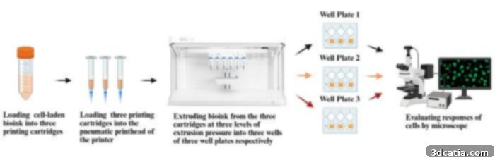
Overview of the experimental design demonstrating the effects of extrusion pressure. (Image Credits: Taieba Tuba Rahman et al).
The work at Texas A&M University aligns with similar advancements in the field of bioprinting. Earlier this year, researchers at McMaster University in Ontario, Canada, developed a bioink that effectively replicates the elasticity and stretchability of native lung tissue. This bioink is particularly well-suited for modeling conditions such as COPD and pulmonary fibrosis, as well as for conducting toxicity and drug-response testing. The ability to mimic the mechanical properties of lung tissue is crucial for creating accurate and physiologically relevant models.
Also this year, the Science & Technology Division of the U.S. Department of Homeland Security, in collaboration with the Wake Forest Institute for Regenerative Medicine, utilized 3D-printed lung tissue to analyze the effects of toxic fumes on human health. This research highlights the potential of 3D-printed lung tissue for assessing the toxicity of environmental pollutants and developing strategies to protect human health.
Collectively, these projects emphasize that biomimetic 3D models are rapidly becoming practical platforms for diagnostics, toxicology, and therapeutic development. The ability to create realistic and functional 3D models of human tissues is transforming the landscape of biomedical research and holds immense promise for improving human health.
To delve deeper into the Texas A&M University study, you can read the original article here.
What are your thoughts on this groundbreaking study? Share your opinions in the comments below or on our LinkedIn and Facebook pages. To stay up-to-date with the latest 3D printing news, subscribe to our free weekly Newsletter. You can also find all our videos on our YouTube channel. For more information on medical and dental 3D printing news, visit our dedicated page HERE.
*Cover Image Credit: Deposit Photos