

Revolutionizing Regenerative Medicine: The Dawn of In-Body 3D Printing with Focused Ultrasound
Imagine a future where a surgeon could repair damaged tissue or even print new organs directly inside your body, without the need for extensive open surgery. While this vision might sound like something plucked from the realm of the most advanced science fiction, groundbreaking research conducted by teams at Duke University and Harvard Medical School suggests it is rapidly moving towards becoming a tangible reality. These visionary researchers have pioneered a novel 3D printing process that leverages the power of focused ultrasound technology in conjunction with a specially formulated, biocompatible ink. This innovative synergy holds immense potential, promising applications that span from accelerating bone healing to precisely repairing delicate heart valves, ultimately paving the way for significantly safer, less invasive, and more effective surgical procedures.
For years, the medical community has recognized and championed the transformative benefits of additive manufacturing within the medical sector. The burgeoning bioprinting field, in particular, has seen remarkable advancements, offering hope for personalized implants, drug testing models, and even organ substitutes. However, translating these advancements into applications *within* the human body has presented significant, often insurmountable, obstacles. The primary challenge lies in the inherent limitations of conventional 3D printing techniques when attempting through-tissue fabrication. Most existing bioprinting processes rely either on extrusion-based methods or various forms of photopolymerization, neither of which is inherently well-suited for the complex and dynamic environment inside a living organism.

This innovative technique utilizes ultrasound-sensitive ink to construct intricate 3D structures at various depths, including through diverse tissues found within the human body, opening new frontiers for in-situ bioprinting.
The limitations of current bioprinting methods for in-body applications are multifaceted. With extrusion-based 3D printing, the difficulty is immediately apparent: how does one introduce and operate a precision printing apparatus deep within the body without resorting to highly invasive surgical incisions? If significant incisions are already necessary, the practical advantages of in-body printing diminish, as external fabrication often becomes a more viable and controlled option. Meanwhile, the array of photopolymerization methods, which utilize light to cure bio-inks, face an equally critical hurdle. The fundamental problem lies in light’s inability to penetrate human skin, organs, and other biological tissues effectively. As light attempts to traverse these dense, multi-layered structures, it undergoes significant scattering and absorption, preventing it from reaching the target area with the necessary precision and intensity. This issue also plagues advanced volumetric printing techniques, which, despite their innovative approach using photopolymerization and transparent inks to create entire structures at once, still rely on light and thus struggle with deep tissue penetration.
Addressing these critical limitations, the research teams have introduced a revolutionary methodology termed deep-penetrating acoustic volumetric printing, or DVAP. This innovative process holds the key to overcoming the inherent challenges of in-body 3D printing. Randy King, Ph.D., a distinguished program director in the Division of Applied Science & Technology at the National Institute of Biomedical Imaging and Bioengineering (NIBIB), eloquently articulates the significance of this development. He explains, “Focused ultrasound has been a cornerstone of medical treatments for decades, demonstrating its unparalleled safety and utility across a broad spectrum of clinical conditions. This potential new application of 3D printing, built upon years of diligent technological advancements, truly has the capacity to lay the groundwork for something previously considered unfathomable: the ability to perform precise 3D ultrasound printing directly through biological tissues.” The implications for regenerative medicine and minimally invasive surgery are profound.
How Does Ultrasound 3D Printing Work? Unveiling the DVAP Mechanism
As Dr. King highlighted, focused ultrasound is far from a newcomer in the landscape of medical technology. Defined by the National Institute of Health as “a non-invasive therapeutic technique that directs ultrasonic waves to a specific location,” it has already established itself as a versatile and effective tool. It is routinely employed in the treatment of various conditions, from addressing liver tumors and uterine fibroids to managing symptoms of neurological disorders like Parkinson’s disease. Its ability to deliver highly localized energy without requiring incisions has made it invaluable. However, this pioneering research marks a pivotal moment, representing the very first time focused ultrasound has been harnessed specifically for the intricate and transformative purpose of medical 3D printing, especially for printing *inside* the body.
The operational principle of DVAP draws parallels with other sophisticated biomedical 3D printing processes, particularly those that employ photo-sensitive inks and targeted light, including advanced volumetric 3D printing. The crucial distinction and innovation in DVAP, however, lies in the development of a unique “sonicated ink,” or “sono-ink,” which possesses an exquisite sensitivity to ultrasound waves rather than light. This ingeniously formulated sono-ink is a composite material, meticulously designed with four distinct components, each playing a vital role in the printing process. Firstly, it contains a specific compound engineered to efficiently absorb ultrasound waves. Secondly, microparticles are integrated to precisely control the ink’s viscosity, ensuring optimal flow and structural integrity during deposition. A polymer provides the foundational structural framework, lending shape and mechanical properties to the printed construct. Finally, a specialized salt is incorporated, which acts as a heat absorber, specifically triggering the solidification process when the localized temperature reaches a critical threshold due to ultrasound absorption. Working in concert, these components enable the precise and controlled printing of biocompatible structures, even when operating through the body’s thick, multi-layered, and often opaque tissues.
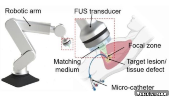
A closer look at how the deep-penetrating acoustic volumetric printing (DVAP) process works, illustrating the precise targeting of ultrasound waves to solidify bio-ink for internal tissue regeneration.
Beyond its core functionality, this sono-ink boasts remarkable adaptability, allowing for tailoring to diverse medical applications. For instance, by integrating bone mineral particles, the ink can be optimized for the regeneration and healing of bone loss, offering a novel solution for orthopedic repair. Furthermore, its mechanical properties can be finely tuned, enabling the creation of structures that are either more durable for long-term support or degradable for temporary scaffolding, depending on the specific physiological needs of a patient and the intended therapeutic outcome. This unparalleled level of flexibility is absolutely critical in advanced medical applications, where patient-specific solutions are paramount. Complementing this versatile ink, the researchers have developed a sophisticated, self-made confocal high-intensity ultrasound printer. This specialized device has been meticulously engineered and adapted to significantly enhance both the speed and resolution of the printing process, ensuring that intricate biological structures can be fabricated with unprecedented precision and efficiency.
The pioneering DVAP process is the brainchild of a collaborative effort between two leading scientists: Y. Shrike Zhang, an associate bioengineer at Brigham and Women’s Hospital and associate professor at Harvard Medical School, and Junjie Yao, an associate professor of biomedical engineering at Duke University. Professor Yao elaborates on the underlying physical principle that powers DVAP: “DVAP relies fundamentally on the sonothermal effect, a phenomenon that occurs when focused sound waves are absorbed by the sono-ink, leading to a highly localized and precise increase in temperature. This localized heating then triggers the hardening and solidification of our specialized ink. Crucially, ultrasound waves possess an extraordinary ability to penetrate biological tissues, reaching depths more than 100 times greater than what can be achieved with light, all while maintaining excellent spatial confinement. This means we can access and precisely print within deep tissues, bones, and internal organs with a high spatial precision that has simply been unreachable using conventional light-based printing methods.” This deep penetration and precise control represent a paradigm shift for in-body manufacturing.
Moreover, the benefits of DVAP extend beyond merely enabling in-body 3D printing through the use of deep-penetrating ultrasound waves. A significant aspect of this breakthrough is the demonstrated compatibility of the process with living biological tissues. Professors Zhang and Yao have already conducted compelling preliminary tests, successfully using the DVAP process to create intricate tissue structures within a pig liver. They have also performed a mock surgical procedure involving a goat heart, further showcasing the technique’s potential. These rigorous experiments have yielded exceptionally promising results, strongly indicating that in the near future, a process like DVAP could realistically be deployed to replace many highly invasive surgical procedures currently used for tissue and organ repair. This could translate to reduced patient trauma, faster recovery times, and significantly improved patient outcomes.
Professor Yao concludes, encapsulating the immense potential of this advancement: “Because our technology allows for precise printing directly through existing tissue layers, it unlocks a vast array of potential applications in both surgery and therapy. These are areas that have traditionally relied on extremely invasive and disruptive methods, often accompanied by significant risks and recovery periods. This work not only represents a monumental leap forward but also opens up an incredibly exciting new avenue within the entire world of 3D printing and regenerative medicine. We are enthusiastically committed to exploring the full potential of this revolutionary tool together, for the benefit of patients worldwide.” For those eager to delve deeper into this transformative research, additional information can be found in the comprehensive press release from the National Institute of Health HERE, or by accessing the full scientific study published in Science HERE.
What are your thoughts on the groundbreaking use of focused ultrasound for medical 3D printing and its implications for future healthcare? We encourage you to share your perspectives in a comment below or join the conversation on our LinkedIn, Facebook, and Twitter pages! Don’t forget to sign up for our free weekly Newsletter here to receive the latest 3D printing news straight to your inbox! You can also find all our engaging videos and in-depth discussions on our YouTube channel.
*All Image Credits: Junjie Yao (Duke University) and Yu Shrike Zhang (Harvard Medical School and Brigham and Women’s Hospital)