

Revolutionizing Childhood Cancer Research: 3D Bioprinting Unlocks Neuroblastoma Secrets and Paves the Way for Advanced Treatments
The fight against childhood cancer is a global imperative, and innovative research is constantly pushing the boundaries of what’s possible. At the forefront of this effort, the INCLIVA Health Research Institute, located within the Hospital Clínico de Valencia in Spain, has made significant strides by harnessing the power of advanced 3D technology. A dedicated research team at INCLIVA has successfully utilized sophisticated 3D bioprinting techniques to gain unprecedented insights into the behavior of aggressive tumors responsible for childhood cancers, specifically focusing on neuroblastoma. This groundbreaking study involved the meticulous application of digital pathology tools to analyze intricate artificial tumor models, providing a more accurate representation of complex biological processes.
The primary objective of this pioneering research was multifaceted: to profoundly understand the dynamics of neuroblastoma cells in terms of their growth patterns, inherent aggressiveness, and critical cell migration capabilities. By creating highly realistic three-dimensional environments that mimic human tissues, researchers can observe these behaviors with a clarity previously unattainable in traditional two-dimensional laboratory settings. Through this ambitious initiative, the INCLIVA Institute aims to leverage the full potential of 3D bioprinting technology not only to enhance existing treatments for childhood cancer but, more importantly, to accelerate the discovery and development of entirely new, more effective therapeutic strategies, ultimately advancing the global fight against this devastating disease.
Understanding Neuroblastoma: A Challenging Childhood Cancer
Neuroblastoma stands as one of the most common solid tumors in infants and young children, often diagnosed before the age of five. This aggressive cancer originates from immature nerve cells, known as neuroblasts, which are typically found in the sympathetic nervous system – a network of nerves that runs through the body and is involved in involuntary functions like heart rate and digestion. Most commonly, neuroblastoma develops in the adrenal glands, located atop the kidneys, but it can also form in nerve tissue in the neck, chest, abdomen, or pelvis. The unpredictable nature of neuroblastoma, ranging from spontaneous regression in some cases to rapid progression and metastasis in others, makes it particularly challenging to treat. Its molecular heterogeneity and tendency to recur even after intensive therapy underscore the urgent need for novel research approaches that can unravel its complex biology and identify vulnerable points for therapeutic intervention.
The Evolving Landscape of Medical 3D Printing: A New Hope
The remarkable advancements in medical 3D printing continue to captivate and inspire, opening new avenues for research and clinical applications across various medical disciplines. We’ve witnessed numerous projects demonstrating the transformative power of this technology. For instance, a recent initiative utilized bioprinting to develop functional pancreas models, offering researchers an unparalleled platform to meticulously study diabetes treatments and disease progression in a highly controlled environment. Similarly, 3D printing is revolutionizing areas from personalized surgical guides and patient-specific implants to prosthetics and sophisticated drug delivery systems. The ability to create intricate, biologically relevant structures with high precision makes additive manufacturing an invaluable tool in the modern medical toolkit. In the context of the INCLIVA study, this cutting-edge technology, stemming from the meticulous doctoral thesis of Ezequiel Monferrer and published in the esteemed International Journal of Molecular Sciences, represents a monumental step forward in leveraging 3D technology to combat the pervasive challenges of childhood cancer.
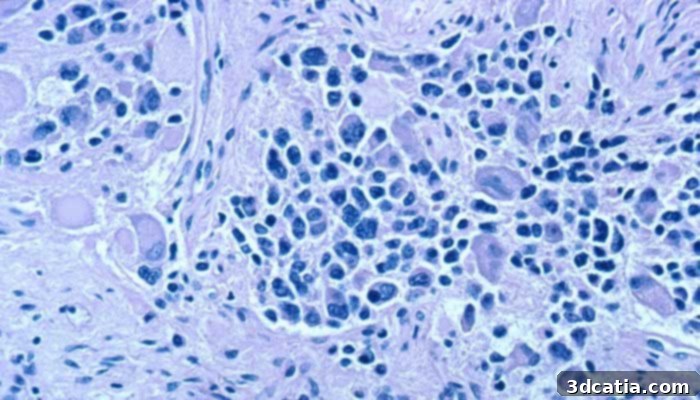
Microscopic view of cells from a nerve ganglion with neuroblastoma.
Pioneering Research: 3D Bioprinting to Combat Childhood Cancer
The core of INCLIVA’s groundbreaking project lies in its innovative approach to understanding tumor biology. Traditional two-dimensional cell cultures, while useful, often fail to accurately replicate the complex cellular interactions and microenvironmental cues present within a living organism. Recognizing this limitation, INCLIVA researchers embarked on a mission to transcend conventional methods by recreating highly realistic 3D models of neuroblastomas. This advanced methodology allows for a far more accurate study of tumor behavior, providing insights that are directly translatable to in vivo conditions.
Within these meticulously engineered 3D structures, neuroblastoma cells were not cultured in isolation. Instead, they were strategically co-cultured with various non-cancerous cells. This crucial step was taken to mimic the intricate tumor microenvironment, recreating the dynamic and often symbiotic relationship between cancerous cells and their surrounding healthy tissues. This interaction is known to significantly influence tumor growth, progression, and resistance to therapy. Ezequiel Monferrer, a key figure in this research, elaborated on the meticulous development process: “Since the project’s inception, we have diligently developed a diverse range of 3D models utilizing three-dimensional bioprinting technology. These models are directly based on patient-derived tumor cells, ensuring their clinical relevance. The structures themselves are meticulously fabricated using biocompatible materials such as gelatin and methacrylated alginate. These materials are chosen for their ability to accurately replicate the complex physical and biochemical properties of natural tumor environments, including varying levels of rigidity and diverse cellular interactions that are fundamental to tumor progression.” The careful selection of these bio-inks and the precision of the bioprinting process allow for the creation of models that accurately reflect the heterogeneity and complexity of actual neuroblastoma tumors.
Key Findings: Unveiling the Tumor’s Secrets
The results emanating from this groundbreaking project have yielded invaluable insights into neuroblastoma’s intricate biology. The research unequivocally indicated that environmental conditions within the 3D bioprinted models profoundly influenced several critical aspects of tumor behavior. Specifically, the study observed significant impacts on cell proliferation rates, the production of vitronectin (an adhesive glycoprotein involved in cell migration and adhesion), and the expression of genes directly linked to cell migration. These findings underscore the paramount importance of the tumor microenvironment in dictating how cancer cells grow, spread, and interact with their surroundings.
Furthermore, a particularly salient discovery was how the strategic presence of non-cancerous cells within these sophisticated 3D bioprinted cultures dramatically modified the behavior of the neuroblastoma tumor cells. This highlights the complex interplay between cancerous cells and their “normal” cellular neighbors, an interaction often overlooked in simpler in vitro models. Understanding these dynamic relationships is crucial for developing therapies that target not just the cancer cells, but also their supportive environment. Dr. Antonio Llombart, Vice-President of the Spanish Association Against Cancer in Valencia, emphasized the critical importance of these findings, stating: “This research is absolutely essential for establishing well-characterized models that accurately mimic various types of neuroblastoma. Such models provide an indispensable platform upon which to perform rigorous preclinical therapeutic trials. This allows us to diligently work on identifying and testing possible treatments specifically designed to reduce the tumor’s aggressiveness, ultimately leading to more effective interventions for young patients.” These realistic 3D models offer a predictive power far superior to traditional methods, enabling researchers to screen drugs more effectively and understand resistance mechanisms before moving to costly and time-consuming in vivo studies.
Photo Credits: Louis Reed/Unsplash
The Broader Horizon of 3D Bioprinting in Healthcare
While the INCLIVA study spotlights neuroblastoma, its implications resonate far beyond this specific cancer. The success of creating such intricate and functional tumor models demonstrates the immense capabilities of 3D bioprinting technology across a multitude of healthcare fields. From drug discovery and toxicology testing to regenerative medicine and personalized surgical planning, 3D printing is transforming how medical research is conducted and how clinical treatments are delivered. The ability to produce ‘organ-on-a-chip’ systems, for example, allows for drug screening that more accurately predicts human response than animal models, reducing development costs and time. For cancer research specifically, these 3D models can serve as unparalleled platforms for testing new chemotherapies, immunotherapies, and targeted drugs in an environment that closely mimics the human body, accelerating the path from laboratory to clinic. They also hold promise for studying drug resistance mechanisms and identifying biomarkers for personalized medicine, tailoring treatments to individual patient needs based on their unique tumor characteristics.
Looking Ahead: Future Directions in the Fight Against Cancer
In conclusion, this landmark study from the INCLIVA Health Research Institute powerfully underscores the immense potential and capabilities of 3D bioprinting technology in addressing some of the most pressing challenges in healthcare, particularly in the critical domain of childhood cancer. By providing researchers with unprecedented tools to observe and manipulate tumor behavior in a highly realistic 3D context, this technology promises to unlock deeper understandings of disease progression and response to therapy. The path forward involves continued refinement of these bioprinted models, integrating more diverse cell types, vascularization, and immune components to make them even more physiologically relevant. The ultimate goal remains to translate these laboratory insights into tangible clinical benefits, delivering an optimal solution that can significantly facilitate and strengthen the global fight against aggressive tumors like neuroblastoma. This research offers a beacon of hope, moving us closer to a future where childhood cancer is not just treatable, but curable, with minimal long-term side effects for our youngest patients.
What are your thoughts on the revolutionary use of 3D printing in the ongoing fight against cancer and other diseases? We’d love to hear your perspective! Please share your insights and comments below, or engage with us on our social media platforms: Facebook, Twitter and LinkedIn. To stay informed about the very latest breakthroughs and innovations in 3D printing, remember to sign up for our free weekly Newsletter here, delivering the most pertinent 3D printing news directly to your inbox!