
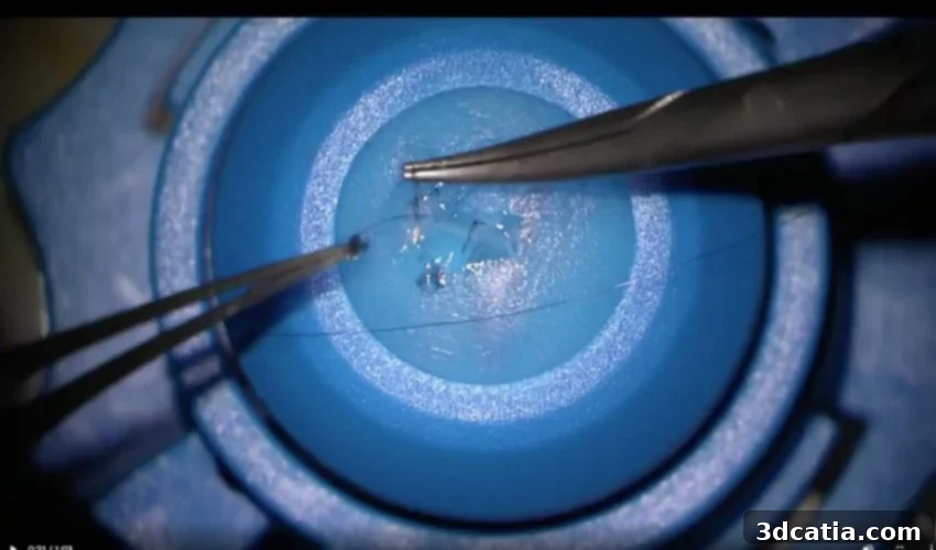
Revolutionizing Ophthalmic Training: How 3D Printed Cornea Models Enhance Surgical Confidence
The field of medicine is continually seeking innovative solutions to enhance patient care, and one technology proving particularly transformative is 3D printing. Often hailed for its utility in various sectors, 3D printing is rapidly becoming indispensable for surgical training and the creation of highly realistic models of complex medical conditions. This is especially true for intricate procedures that are challenging to practice outside the demanding environment of an operating theater. Among these, delicate eye surgeries stand out, where precision is paramount and the margin for error is virtually non-existent. Specifically, the treatment of trauma to the cornea – the transparent, outermost layer of the eye covering the iris and pupil – presents a significant challenge for surgeons, necessitating extensive and meticulous training.
Recognizing this critical need, a pioneering team of researchers at King’s College Hospital in the UK has embarked on an innovative project, developing a groundbreaking trauma simulation model using advanced 3D printing technology. Their preliminary data is highly encouraging, suggesting that these sophisticated models offer a valuable and effective option for significantly improving the confidence and proficiency of surgeons and clinical fellows tasked with treating eye trauma. This development marks a pivotal step forward in medical education, providing a much-needed bridge between theoretical knowledge and practical expertise in a controlled, safe environment.
Developing Realistic Cornea Trauma Models
The creation of these highly accurate cornea models involved a meticulous design process, leveraging cutting-edge CAD (Computer-Aided Design) software. The researchers utilized Fusion 360, a powerful and versatile software known for its robust modeling capabilities. The designs were not merely theoretical constructs; they were meticulously informed and based on real-life clinical cases encountered at King’s College Hospital, ensuring that the models accurately reflected the complexities and variations of actual corneal trauma. This empirical foundation is crucial for developing training tools that truly prepare surgeons for diverse scenarios.
Once the digital designs were finalized, the team turned to advanced additive manufacturing for production. They specifically employed the Stratasys J850, a high-fidelity multi-material 3D printer renowned for its ability to produce highly detailed and color-accurate models. The models were printed using a specialized photopolymer material, carefully selected for its ability to mimic the tactile properties and visual characteristics of real corneal tissue. This material choice is critical, as it allows trainees to experience the haptic feedback and visual cues that are essential for developing fine motor skills and accurate surgical techniques. The result was a set of incredibly realistic cornea models, complete with intricate details representing various forms of traumatic injury.
Impact on Surgical Training and Confidence
To assess the efficacy of their 3D printed models, the King’s College Hospital team conducted a comprehensive simulation course. This intensive training program involved a group of 14 experienced ophthalmologists and 5 clinical fellows, all of whom regularly face the challenges of eye trauma. Participants engaged in hands-on practice, using the 3D printed models to hone their assessment and suturing skills under simulated operating conditions. The controlled environment allowed them to experiment, make mistakes, and refine their techniques without any risk to patients.
Following the training course, participants completed a detailed questionnaire designed to gauge their perceptions and confidence levels. The results were overwhelmingly positive: every single participant reported feeling significantly more confident with both the assessment and the intricate suturing processes required for corneal repair compared to their confidence levels before the simulation. This unanimous positive feedback underscores the profound impact these 3D printed models can have on surgical readiness. Crucially, these innovative 3D models represent a highly effective and ethically sound alternative to the current, often limited, options for surgical training, which typically involve the use of animal or cadaver eyes. The ethical implications and practical challenges associated with sourcing and using biological materials make the 3D printed alternatives a superior choice.

Beyond surgical models, 3D printing has demonstrated its versatility in creating prosthetic eyes, such as this example for patient Steve Verzey in November 2021. This highlights the broader application of the technology in restoring function and aesthetics for patients. (Photo credit: Moorfields Eye Hospital)
The Broader Advantages of 3D Printing in Medical Training
According to the King’s College researchers, the integration of 3D printing into medical training offers a myriad of benefits that extend beyond mere confidence building. One significant advantage is the drastically shorter lead time for producing training models. Unlike traditional methods that can involve lengthy procurement processes or complex fabrication, 3D printing allows for rapid prototyping and on-demand production. This agility means that new models, perhaps reflecting rare or unique trauma cases, can be designed and printed quickly, keeping training materials current and highly relevant. Furthermore, the ability to mass-produce these models within a hospital setting is a game-changer for skills training programs.
This capability empowers doctors and medical students to dedicate valuable time to hone their skills outside the high-pressure environment of an operating theater. In a live surgery, the stakes are incredibly high, and opportunities for experimental practice are naturally limited. 3D printed models provide a safe, repeatable, and low-stress environment where practitioners can refine their techniques, understand anatomical variations, and manage complications without the direct supervision of senior staff always being immediately necessary. While training on real patients remains an absolutely essential component of acquiring the nuanced technical skills required in surgery, practice on highly realistic models offers a foundational layer of competence and comfort. It bridges the gap between theoretical knowledge and practical application, ensuring that when trainees do enter the operating room, they are better prepared, more efficient, and ultimately safer for patients.
3D Printing: A Versatile Tool Across Healthcare
The application of 3D printing in a hospital context to aid learning and treatment is not a novel concept, though its capabilities continue to expand. The King’s College Hospital initiative is one more testament to its burgeoning role. For instance, in 2021, we reported on Seattle Children’s Hospital’s innovative use of Stratasys technology to create highly detailed anatomical models for surgical preparation. These models allowed surgeons to practice complex procedures on patient-specific anatomies, greatly reducing operative time and improving outcomes for challenging pediatric cases.
Beyond training and planning, 3D printing has also made significant inroads into the treatments themselves. A particularly compelling recent example comes from IUCT Oncopole (University Cancer Institute of Toulouse), where surgeons successfully bioprinted a nose graft. This remarkable procedure involved growing the bioprinted tissue on a patient’s forearm before it was surgically implanted onto her face during reconstructive surgery. This showcases the incredible potential of bioprinting to create living tissues and organs, promising a future where customized biological structures can replace damaged or diseased ones. From highly complex surgical guides and patient-specific implants to advanced prosthetics and the pioneering field of bioprinting, additive manufacturing is consistently pushing the boundaries of what is possible in modern healthcare.
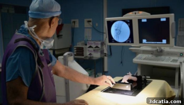
3D printing technology offers the ability to create highly realistic and patient-specific surgical models of organs, aiding in complex procedure planning and training. (Photo credit: Springwise/Biomedex)
The Future of Ophthalmic Surgery and Medical Education
The research conducted by King’s College Hospital, published in “Eye,” the official journal of The Royal College of Ophthalmologists, provides robust evidence for the immediate benefits of 3D printed models in ophthalmic training. The study is a testament to the increasing sophistication and accessibility of 3D printing technology, making advanced medical training more democratic and effective. These advancements not only enhance the skills of individual surgeons but also contribute to a broader improvement in patient safety and surgical outcomes across the healthcare system.
As 3D printing technology continues to evolve, we can anticipate even more sophisticated and hyper-realistic models, potentially incorporating dynamic elements or varying tissue properties to simulate complex physiological responses. This ongoing innovation promises to further refine surgical education, offering unprecedented opportunities for learning and skill acquisition. The integration of such technologies into medical curricula worldwide could fundamentally transform how future generations of medical professionals are trained, moving towards a more hands-on, personalized, and ethically sound approach to skill development. The full study detailing this significant contribution to medical science can be accessed via the provided link HERE.
What are your thoughts on the use of 3D printing for eye trauma modeling and its broader implications for medical training? We invite you to share your insights and comments below or engage with our community on our social media platforms. Find us on Linkedin, Facebook, and Twitter! To stay updated with the very latest developments and news in the exciting world of 3D printing, don’t forget to sign up for our free weekly Newsletter here, delivered straight to your inbox. You can also explore all our informative videos and engaging content on our dedicated YouTube channel, offering a visual journey through the innovations of additive manufacturing.