
Revolutionizing Cardiovascular Treatment: How 3D Printed Vascular Grafts with TMP Combat Thrombosis and Aneurysms
When considering the world’s most dangerous diseases, many minds immediately turn to cancer. However, the sobering reality is that cardiovascular disease (CVD) holds this unfortunate distinction, standing as the leading cause of death globally. Even after successful interventions, patients often face debilitating complications, including thrombosis—the formation of blood clots that can block vessels—and aneurysmal dilation, a dangerous widening of arteries post-surgery. These challenges underscore the urgent need for more effective and safer treatment modalities for a variety of cardiovascular conditions.
Fortunately, a groundbreaking solution may be on the horizon. A dedicated research team, collaborating between Donghua University and Shanghai Jiao Tong University, has recently published a pivotal study. Their innovative work introduces a novel approach: 3D printed electrospun vascular grafts infused with tetramethylpyrazine (TMP). TMP, a naturally occurring chemical compound found in traditional Japanese nattō and fermented cocoa beans, has demonstrated remarkable therapeutic potential. The study’s findings suggest that these specially engineered grafts could significantly improve cardiovascular disease treatments by effectively reducing thrombosis and preventing aneurysmal dilation following surgical procedures.
The global impact of cardiovascular diseases is staggering. According to the British Heart Foundation, an estimated 620 million people worldwide live with some form of CVD, representing approximately 1 in every 13 individuals. This makes it not only a widespread condition but also the primary cause of mortality globally, accounting for at least a third of all deaths. To put this into perspective, cancers are responsible for roughly one-in-five deaths. A common and critical treatment for severe CVD, particularly due to conditions like stenosis (the total narrowing of arteries in the heart), involves vessel replacement. However, current standard vascular grafts, while life-saving, are far from perfect. They frequently encounter significant challenges such as thrombosis, restenosis (re-narrowing of the vessel), infection, and inadequate mechanical properties, which can lead to complications and the need for repeat interventions. This is precisely where the innovative 3D printed electrospun vascular grafts are poised to make a profound and positive difference in patient outcomes.
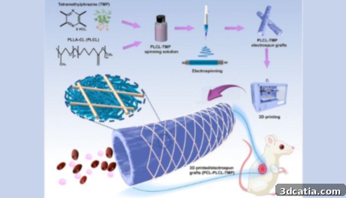
An illustration of the 3D printed electrospun vascular graft filled with TMP (image credits: Burns & Trauma)
Addressing Cardiovascular Disease with Advanced 3D Printed Vascular Grafts
The development of these next-generation vascular grafts represents a significant leap forward in medical technology, combining the precision of additive manufacturing with advanced materials science. The grafts were meticulously fabricated through a hybrid process that integrates electrospinning with a specialized 3D printing technique. Electrospinning is an electrohydrodynamic process renowned for its ability to produce extremely fine polymer fibers, often in the nanoscale, which closely mimic the natural extracellular matrix found in biological tissues. This technique is crucial for creating scaffolds that promote cell adhesion and growth, essential for tissue regeneration.
The innovative design features a sophisticated dual-layer structure, carefully engineered to provide optimal stability, flexibility, and biological compatibility. The inner layer, which comes into direct contact with blood, is composed of electrospun poly (L-lactic-co-caprolactone) (PLCL) nanofibers. PLCL is a biocompatible and biodegradable polymer known for its excellent elasticity, which is vital for mimicking the pulsatile nature of natural blood vessels. The nanofibrous structure of this layer is key to promoting endothelial cell growth and minimizing platelet adhesion, thereby directly addressing the risk of thrombosis. The outer layer provides structural integrity and mechanical strength, being constructed from 3D printed polycaprolactone (PCL) microfibers. PCL is another biodegradable polymer, chosen for its robust mechanical properties and slow degradation rate, ensuring long-term support for the graft. The strategic combination of these two layers and materials ensures that the graft is both mechanically resilient and biologically responsive, capable of withstanding the dynamic forces within the circulatory system while supporting tissue regeneration.
A critical enhancement to these grafts is the incorporation of tetramethylpyrazine (TMP). Derived from the traditional Chinese medicine *Ligusticum chuanxiong*, TMP is celebrated for its potent antiplatelet and anticoagulant properties. These properties are invaluable in the context of vascular grafts, as they directly combat the formation of blood clots, which is a primary cause of graft failure and post-surgical complications like acute thrombosis. By integrating TMP directly into the graft material, the researchers aim to provide a localized and sustained release of the compound, enhancing the safety and longevity of the implant without the systemic side effects often associated with orally administered anticoagulants. This innovative drug-eluting approach promises to significantly improve the early stages of graft integration and reduce the likelihood of life-threatening thrombotic events.
The research yielded highly encouraging results across both in vitro and in vivo studies. In vitro tests, conducted under laboratory conditions, unequivocally demonstrated that the 3D printed grafts were remarkably effective in reducing platelet adhesion. This finding is of paramount importance in the fight against thrombosis, as platelet aggregation is the initial step in clot formation. Furthermore, these tests confirmed excellent compatibility with human cells, indicating a low risk of immunological rejection and promoting cellular integration—crucial factors for the long-term success of any implantable medical device. The grafts exhibited a favorable environment for cellular growth and function, essential for the natural remodeling and healing processes within the body.
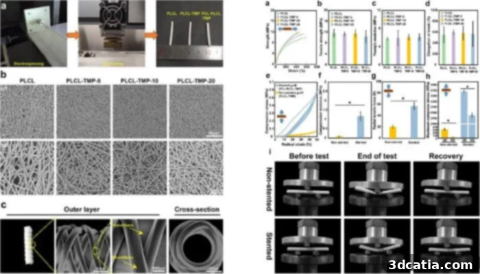
(Left) A closer look at the preparation of the graphs; (right) the results from different tests (image credits: Burns & Trauma)
The success was further validated through comprehensive in vivo experiments conducted in a rat model. Here, the animals underwent surgical procedures where sections of their abdominal aortas were replaced with the innovative 3D printed grafts. Over a remarkable period of six months, the grafts demonstrated exceptional biocompatibility, seamlessly integrating with the surrounding tissues without adverse reactions. Crucially, they also maintained robust mechanical strength and structural integrity under physiological loads, proving their durability in a dynamic biological environment. Most importantly, the results were overwhelmingly positive regarding critical post-surgical complications: neither acute thrombosis nor significant aneurysmal dilatation was detected in any of the treated rats. These findings are monumental, showcasing the immense potential of these grafts for dramatically improving outcomes in patients who require vascular replacement or reconstruction. The ability to prevent both immediate clotting and long-term structural weakening positions this technology as a transformative advancement in vascular tissue engineering.
Dr. Hongbing Gu, a lead researcher involved in this pioneering study, underscored the significance of their achievements, concluding, “This study marks a significant advancement in vascular tissue engineering. The strategic combination of electrospinning and 3D printing, coupled with the targeted incorporation of TMP, has resulted in a vascular graft that not only meets stringent mechanical requirements but also exhibits exceptional blood compatibility. These compelling findings pave the way for exciting future clinical applications, promising a new era of safer and more effective treatments for cardiovascular patients.” The immediate next steps for the research team involve expanding their studies to large animal models, a crucial phase to further validate the long-term efficacy, safety, and scalability of these grafts before moving towards human clinical trials. In the interim, for those eager to delve deeper into the technical specifics and comprehensive results of this groundbreaking work, the full published paper is accessible HERE. This research not only offers hope for millions suffering from cardiovascular diseases but also highlights the immense potential of integrating advanced manufacturing techniques like 3D printing with innovative biomaterials and therapeutic compounds to redefine modern medicine.
What are your thoughts on this revolutionary development in 3D printed vascular grafts? We invite you to share your insights and comments below, or engage with us on our LinkedIn, Facebook, and Twitter pages! Don’t miss out on the latest advancements and breaking news in additive manufacturing by signing up for our free weekly newsletter here, delivered straight to your inbox. For a visual dive into the world of 3D printing, explore all our videos on our YouTube channel, where innovation comes to life.