

University of Minnesota Pioneers 3D Printed Stem Cell Scaffolds for Spinal Cord Injury Repair
Spinal cord injuries (SCIs) represent one of the most debilitating conditions faced by individuals globally, profoundly impacting their quality of life. In the United States alone, an estimated 300,000 people live with the consequences of an SCI, with approximately 18,000 new cases occurring annually. These life-altering injuries can result from various traumas, such as accidents, falls, or sports-related incidents, as well as from certain medical conditions. Statistically, men are disproportionately affected by SCIs. The devastating impact of these injuries is largely due to the central nervous system’s limited capacity for self-repair. Once nerve cells are destroyed, and nerve fibers are severed at the injury site, the body struggles to regenerate functional connections, often leading to permanent paralysis, loss of sensation, and significant impairments in motor function.
For decades, medical science has grappled with the complexity of spinal cord injuries. Current treatments primarily focus on stabilization, surgical intervention to decompress the spinal cord, and extensive rehabilitation to manage symptoms and maximize remaining function. However, no existing treatment option has been able to fully restore lost body functions, leaving a significant unmet medical need. The core challenge lies in overcoming the destruction of neural tissue and the formation of glial scars, which inhibit axonal regeneration and functional reconnection.
However, a groundbreaking development from researchers at the University of Minnesota Twin Cities offers a beacon of hope in this challenging field. For the first time, their innovative approach synergizes three cutting-edge technologies: advanced 3D printing, potent stem cell therapy, and sophisticated lab-grown tissue engineering. This pioneering method opens unprecedented possibilities for not only repairing spinal cord injuries but also actively promoting functional recovery in a way previously considered beyond reach. This represents a significant leap forward in regenerative medicine, potentially transforming the lives of countless individuals living with SCI.
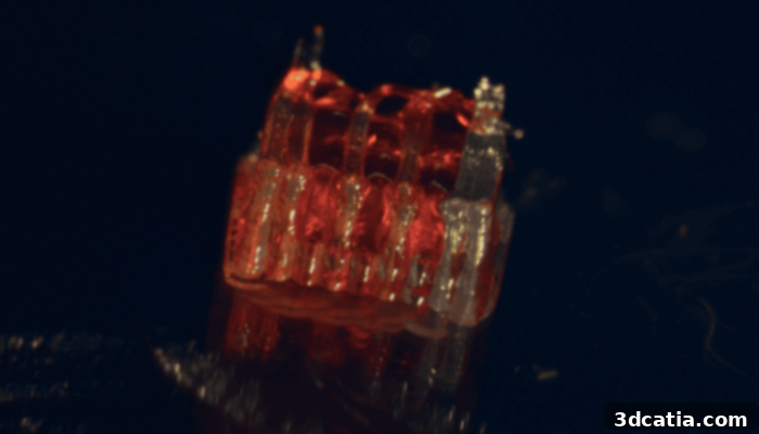
Photo Credit: University of Minnesota
3D-Printed Scaffolds: Engineering a Path for Spinal Cord Regeneration
The core of this revolutionary technique revolves around the meticulous fabrication of a 3D-printed structure, which the researchers aptly named an “organoid scaffold.” This meticulously engineered framework is specifically designed to host and nurture lab-grown tissues, providing a supportive environment for neural regeneration. The scaffold’s intricate design incorporates tiny, microscopic channels that serve a critical purpose: they act as a guide, directing the precise growth of new nerve fibers. Into these channels, spinal cord–specific neural progenitor cells are carefully introduced.
The Power of Neural Progenitor Cells
These specialized cells are not just any cells; they are derived from human adult stem cells, endowing them with extraordinary capabilities. Neural progenitor cells possess the unique ability to multiply rapidly and, more importantly, to differentiate into various types of mature nerve cells, including neurons and glial cells. This remarkable plasticity makes them ideal candidates for regenerating damaged neural pathways. Once embedded within the 3D-printed scaffold, these cells embark on a journey of growth and differentiation, guided by the scaffold’s structure.
Guebum Han, a former postdoctoral researcher in mechanical engineering at the University of Minnesota and the lead author of the study, elaborates on the scaffold’s crucial role: “We use the 3D printed channels of the scaffold to direct the growth of the stem cells, which ensures the new nerve fibers grow in the desired way.” This directed growth is paramount, as uncontrolled or disorganized nerve growth can lead to further complications or ineffective recovery. The scaffold thus acts as a precise architectural blueprint, ensuring that the regenerating nerve fibers extend along a predetermined path, fostering optimal integration with existing neural circuits.
Han further explains the innovative functional outcome: “This method creates a relay system that, when placed in the spinal cord, bypasses the damaged area.” This “relay system” is a critical conceptual breakthrough. Instead of attempting to repair the exact point of injury, which is often riddled with scar tissue, the scaffold creates a bypass—a new functional bridge that allows neural signals to circumvent the lesion. This essentially re-establishes communication pathways that were previously severed, offering a fresh conduit for nerve impulses.
Preclinical Success: Restoring Motor Functions in Rats
To validate their approach, the research team conducted a series of experiments, implanting these innovative scaffolds into rats suffering from severe spinal cord injuries. The results were profoundly encouraging. Post-implantation, the neural progenitor cells within the scaffold successfully transformed into mature neurons. These newly formed neurons then began to extend nerve fibers in both rostral (towards the head) and caudal (towards the tail) directions, actively forming new connections with the rats’ existing neural circuits. Over a period of time, these new cells and their connections demonstrated remarkable integration into the host spinal cord tissue, becoming a seamless part of the biological network.
The ultimate measure of success for any SCI treatment lies in functional recovery. In this regard, the University of Minnesota team’s findings were particularly promising. The rats that received the scaffold implants were observed to regain certain motor functions that had been lost due to their severe spinal cord injuries. This recovery, while not yet full restoration, represents a monumental step forward, providing compelling evidence that this novel combination of 3D printing, stem cell therapy, and tissue engineering holds genuine potential for therapeutic intervention in humans.
Ann Parr, a distinguished professor of neurosurgery at the University of Minnesota, underscores the significance of these findings, stating, “Regenerative medicine has brought about a new era in spinal cord injury research.” Her statement highlights the paradigm shift this research represents—moving beyond merely managing symptoms to actively regenerating and restoring lost neurological function. While acknowledging that this research is still in its early stages and considerable work remains, she emphasizes that it represents a powerful new source of hope for individuals affected by such devastating injuries.
The Future of Spinal Cord Injury Treatment: Scaling Up and Clinical Translation
The success demonstrated in preclinical rat models lays a robust foundation for future development. The immediate next steps for the University of Minnesota team involve scaling up the production of these complex 3D-printed organoid scaffolds. This will necessitate optimizing manufacturing processes to ensure consistency, reproducibility, and the capacity for larger-scale trials. Furthermore, continued development will focus on refining the scaffold design, cell types, and implantation techniques to maximize efficacy and safety. The ultimate goal is to translate this innovative technology into future medical applications, leading towards human clinical trials.
The journey from promising preclinical results to a viable human therapy is long and complex, fraught with regulatory hurdles, extensive safety testing, and the challenges of large-animal models. However, the University of Minnesota’s work is not only advancing the treatment of spinal cord injuries but also pushing the boundaries of what is possible in regenerative medicine. This research showcases the incredible potential of merging advanced manufacturing techniques like 3D printing with biological therapies, offering personalized and highly effective solutions to previously intractable medical conditions.
The broader implications of this research extend beyond spinal cord injuries. The principles developed here could potentially be adapted for repairing other types of neural damage, such as those resulting from stroke, traumatic brain injury, or neurodegenerative diseases. As 3D bioprinting technology continues to evolve, its capacity to create intricate biological structures with unprecedented precision will undoubtedly unlock further advancements in tissue engineering and regenerative medicine, bringing us closer to a future where functional regeneration is a reality for many more patients.
This pioneering work from the University of Minnesota offers a powerful testament to human ingenuity and the relentless pursuit of scientific breakthroughs. It instills genuine optimism that the era of permanent paralysis from spinal cord injuries may one day become a relic of the past, replaced by effective, regenerative treatments that restore hope and function to countless lives.
What do you think of these recent developments from the University of Minnesota? Let us know in a comment below or on our LinkedIn or Facebook pages! Plus, don’t forget to sign up for our free weekly Newsletter to get the latest 3D printing news straight to your inbox. You can also find all our videos on our YouTube channel. Interested in more medical and dental 3D printing news? Visit our dedicated page HERE.
*Cover Photo Credit: Motion Array