
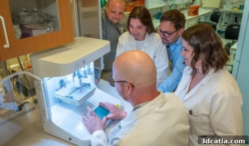
Revolutionizing Burn Care: 3D Bioprinting Living Skin for Advanced Regeneration
The skin, our body’s largest organ, acts as an indispensable shield, protecting us from infections, regulating temperature, and mediating sensory input. When this vital barrier is compromised by severe burns, the consequences can be catastrophic, posing immediate threats to life and long-term challenges for recovery. Traditional burn treatments often involve transplanting thin layers of the epidermis, the outermost skin layer, derived from a single cell type. While these procedures are often lifesaving, they frequently lead to significant scarring, functional limitations, and an aesthetic outcome that can profoundly impact a patient’s quality of life. The need for more effective, regenerative solutions in burn treatment is pressing. In a promising stride forward, researchers from the Center for Disaster Medicine and Traumatology, in collaboration with Linköping University, have developed an innovative gel containing living cells. This groundbreaking bioink is designed to be 3D printed into fully functional skin transplants, offering a new horizon of hope for severe burn victims seeking comprehensive skin regeneration.
Beneath the protective epidermis lies the dermis, a thicker, more intricate layer crucial for the skin’s strength, elasticity, and overall function. This complex tissue houses an extensive network of blood vessels, nerve endings, hair follicles, and sweat glands, all essential for healthy skin. Regenerating this deeper layer is paramount for achieving functional recovery and minimizing scarring in burn injuries. However, transplanting the dermis directly from another part of the patient’s body is rarely a viable option. Such a procedure would invariably create a secondary wound as large and debilitating as the original injury, introducing additional risks, pain, and recovery challenges. Recognizing this significant hurdle, the research team embarked on a mission to engineer a novel solution: living skin tissue that could effectively replace and regenerate the dermis without creating new donor site morbidities. Their innovative approach focuses on leveraging the power of 3D bioprinting and advanced bioink formulations to create a new paradigm in dermal reconstruction, addressing the critical shortcomings of existing methods and paving the way for superior long-term outcomes for burn patients.
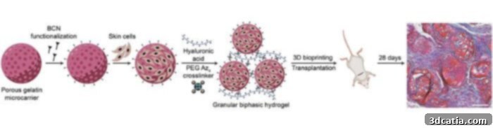
A graphical abstraction of the study illustrating the innovative process. (Image credits: R. Shamasha et al.)
Innovating the Core: Development of a Specialized Bioink for Dermal Regeneration
The success of creating functional 3D bioprinted skin hinges on the development of an effective “bioink”—a printable material containing living cells. For dermal regeneration, the primary cell type targeted by the Linköping University team was fibroblasts. These connective tissue cells are abundant in the dermis and play a pivotal role in synthesizing the extracellular matrix, which provides structural support to tissues. Fibroblasts are particularly advantageous for tissue engineering due to their ease of harvesting from a patient’s own body through a minimal biopsy and their robust capacity for proliferation and growth in a laboratory setting. Furthermore, these versatile cells possess the remarkable ability to mature into other specialized cell types as needed, making them ideal candidates for complex tissue reconstruction.
Initially, the researchers cultivated these crucial fibroblasts within a scaffold composed of tiny, porous gelatin beads. Gelatin, derived from collagen, is highly biocompatible and provides an excellent environment for cell growth. However, this initial formulation presented a significant challenge: the resulting cell-laden liquid lacked sufficient viscosity and structural integrity to remain in place when applied to a wound, or to be precisely 3D printed. To overcome this critical rheological problem, the team devised an ingenious solution. They blended the gelatin beads, with their embedded fibroblasts, with a hyaluronic acid-based gel. Hyaluronic acid is a naturally occurring polysaccharide known for its excellent biocompatibility, biodegradability, and ability to retain moisture, making it a common component in many biomedical applications. The combination of these two components resulted in a novel material with a remarkable property: it exhibits shear-thinning behavior. This means it behaves like a liquid when subjected to mechanical stress, such as pressure from a syringe or a bioprinter nozzle, but quickly reverts to a gel-like state once the stress is removed.
Explaining the unique properties of this innovative substance, Daniel Aili, a distinguished professor of molecular physics at Linköping University and co-lead of the pivotal study, stated, “You can use a syringe to apply it to a wound, for example, and once applied, it becomes gel-like again, effectively staying where it needs to be. This remarkable shear-thinning property also makes it perfectly suitable for precise 3D printing of the gel with the living cells intricately embedded within its structure.” This dual capability – both syringe application and 3D bioprinting – offers significant versatility for future clinical translation, allowing for tailored approaches depending on the burn injury’s characteristics and location.

Scanning electron microscope (SEM) imaging provides a detailed look at the bioprinted cells. (Image credits: R. Shamasha et al.)
In the current pre-clinical study, the research team successfully utilized an extrusion bioprinter to create small, disc-shaped constructs, or “pucks,” from their novel bioink. These bioprinted dermal equivalents, containing the living fibroblasts, were then strategically placed under the skin of mice to evaluate their viability and regenerative potential in a living system. The results of this crucial in-vivo experiment have been exceptionally promising, demonstrating the immense potential of this advanced bioprinting technology. The overarching vision is to enable the growth of a patient’s own cells from a minimal skin biopsy in the lab. These lab-grown, autologous cells could then be incorporated into the bioink and precisely 3D printed into a custom-fit graft, ready for direct application to a severe burn wound. This personalized approach dramatically reduces the risk of immune rejection, a common complication with allogeneic (donor) grafts, and facilitates seamless integration with the patient’s existing tissues.
Johan Junker, a leading researcher at the Swedish Center for Disaster Medicine and Traumatology, a docent in plastic surgery at Linköping University, and co-lead of this pioneering research, enthusiastically commented on the study’s significant outcomes. “We clearly observe that the cells within the bioprinted grafts not only survive but also actively produce various essential substances that are crucial for creating new, functional dermis,” he reported. He further elaborated on a particularly critical finding: “In addition, we detected the formation of new blood vessels within the grafts, which is absolutely vital for the tissue to survive, thrive, and integrate effectively within the body. This vascularization is a key indicator of successful tissue regeneration. We find this innovative material and its regenerative capabilities incredibly promising for the future of burn care.” The ability to promote vascularization is a major breakthrough, as poor blood supply is a common reason for graft failure and limits the thickness of skin constructs that can be successfully transplanted.
3D Printing: A Transformative Force in Modern Burn Treatment
The Linköping University study represents a significant milestone within a broader and accelerating trend of scientific endeavors aimed at fundamentally improving burn treatment through the innovative application of 3D printing technologies. This burgeoning field, often termed regenerative medicine and tissue engineering, promises to overcome many limitations of conventional approaches. Earlier this year, for example, the esteemed Centre for Burn Research at the Hamilton Health Sciences (HHS) hospital network in Canada initiated a groundbreaking clinical trial. This trial is investigating the efficacy of a bioink specifically formulated from a patient’s own burned skin cells, highlighting the move towards highly personalized and autologous solutions in burn care. Beyond direct tissue regeneration, 3D printing is also revolutionizing other aspects of wound management and patient support.
Numerous research groups globally have successfully developed and tested a variety of 3D printed devices designed to enhance wound treatment and patient recovery. These innovations include specialized wound dressings, which can be custom-printed to perfectly fit irregular wound shapes, incorporating therapeutic agents for targeted drug delivery and optimized healing environments. Furthermore, 3D printing has been instrumental in creating custom facial orthoses and prosthetics, offering crucial support for patients, particularly children, recovering from severe facial burns. These devices provide support during healing, help manage scarring, and improve aesthetic outcomes. The integration of advanced manufacturing techniques like 3D printing across the entire spectrum of burn care, from acute treatment to long-term rehabilitation, underscores its potential to transform patient outcomes. For those interested in delving deeper into the specifics of the Linköping University study, additional information can be found by reading their comprehensive article or by accessing the full scientific study published in the esteemed journal Advanced Healthcare Materials. This continuous innovation highlights a promising future where severe burn injuries are treated not just to survive, but to truly thrive with regenerated, functional skin.
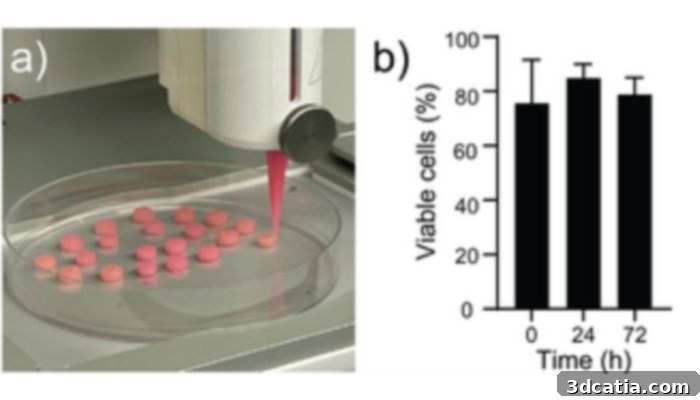
An illustrative diagram of the bioprinting process (a) and a graph demonstrating cell viability, expressed as the percentage of live cells relative to the total number of cells within each construct (b). (Image credits: R. Shamasha et al.)
The development of 3D bioprinted living skin represents a significant leap forward in regenerative medicine, promising to transform the prognosis and quality of life for individuals suffering from severe burn injuries. By enabling the creation of functional dermal tissue with integrated vascularization, this research offers a pathway to not only save lives but also significantly reduce scarring and restore the crucial functions of healthy skin. This innovative approach moves us closer to a future where personalized, regenerative therapies are standard practice, offering renewed hope and improved long-term outcomes for patients worldwide. What are your thoughts on this incredible advancement in 3D bioprinted skin and its potential to revolutionize burn treatment? We invite you to share your insights in a comment below or engage with our community on our LinkedIn or Facebook pages! Don’t miss out on the latest advancements in additive manufacturing—sign up for our free weekly Newsletter to receive the most current 3D printing news directly in your inbox. You can also explore all our informative videos on our dedicated YouTube channel. For more updates and detailed articles on medical and dental 3D printing news, please visit our specialized page HERE.
*Cover Photo Credit: Magnus Johansson