
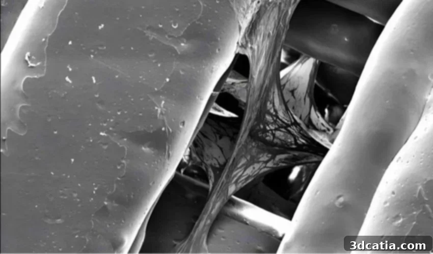
3D Printing Bone Models: Revolutionizing Cancer Research and Reducing Animal Testing
The persistent ethical concerns surrounding animal experimentation, particularly for medical research, have long spurred the scientific community to seek more humane and effective alternatives. In a groundbreaking initiative, a dedicated team of scientists at the prestigious Henry Royce Institute is pioneering the use of advanced 3D printing technology to construct intricate bone structures. This innovative approach aims to create highly realistic microenvironments for the meticulous study of various forms of cancer, especially focusing on how cancer cells interact with and metastasize within bone tissue. By replicating the complex physiological conditions of natural bone, this method significantly reduces the reliance on animal trials, while simultaneously empowering researchers to accelerate and expand the scope of their experiments. The implications are profound, leading us to consider: is this paradigm-shifting practice poised for global democratization, fundamentally altering the landscape of cancer research and drug discovery?
Indeed, the integration of 3D technologies within the medical field is not a novel concept; it has consistently proven instrumental in conducting trials and fostering a deeper understanding of numerous pathologies. From creating patient-specific anatomical models for surgical planning to developing sophisticated prosthetics and implants, additive manufacturing has demonstrated unparalleled versatility. These technologies facilitate the precise creation of tailor-made structures using a diverse array of biocompatible materials, which are crucial for cultivating various cell types under controlled conditions. Furthermore, such structures can be rapidly designed and fabricated to meet specific research or clinical needs, often with remarkably short lead times. The inherent advantages of additive manufacturing in accelerating innovation and enhancing research capabilities are increasingly recognized, and the transformative work being undertaken by the Henry Royce Institute stands as a compelling testament to its enduring potential.
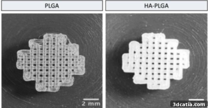
3D printed bone structures mimic natural bone environment for cancer cell study (Photo credits: Fatih Eroglu)
The project at the Henry Royce Institute specifically focuses on 3D printing intricate bone structures designed to serve as meticulously engineered “homes” for cancer cells. This innovative approach is particularly targeted at understanding the complex mechanisms of breast cancer metastasis, a devastating aspect of the disease where cancer cells spread from the primary tumor to distant sites, frequently invading bone tissue. The primary objective is to gain unprecedented insights into how these aggressive cancer cells propagate, interact, and survive within an environment that closely mimics the physiological complexities of living bone. To achieve this high level of realism, the research team employs a sophisticated combination of two key biomaterials: PLGA and HA-PLGA. PLGA (Poly(lactic-co-glycolic acid)) is a well-established, biodegradable synthetic polymer widely recognized for its biocompatibility and tunable degradation rates, forming the foundational matrix of the bone model. The second material, HA-PLGA, is a composite blend that integrates PLGA with hydroxyapatite (HA). Hydroxyapatite is a naturally occurring mineral that constitutes the main inorganic component of bone and teeth, providing bone with its rigidity and strength. By incorporating HA, the researchers are able to engineer a more biologically authentic and mechanically relevant model, ensuring that the cellular environment is as close as possible to natural bone tissue, thereby providing more accurate and reliable data for cancer research.
The choice of fused deposition modeling (FDM) as the preferred 3D printing technology for this critical work highlights a strategic balance between precision and cost-effectiveness. FDM, a highly accessible and widely used additive manufacturing process, operates by extruding heated thermoplastic material layer by layer to build a three-dimensional object. This method allowed the Henry Royce Institute team to significantly curtail production costs. According to the researchers, the FDM 3D printer utilized for this project cost less than 1,000 euros, representing a substantial saving compared to the thousands of euros typically required for specialized 3D bioprinters. This economic accessibility is a key factor in making advanced tissue engineering more widely available to research institutions globally. The FDM technology is employed to precisely print intricate scaffolds, which act as architectural frameworks designed to promote robust cell culture and facilitate the study of cancer cell proliferation and interaction within a simulated bone matrix. Specifically, the researchers introduce bone marrow mesenchymal stem cells (BMSCs) onto these scaffolds. BMSCs are pluripotent stem cells renowned for their ability to differentiate into a variety of cell types, including osteoblasts (bone-forming cells). By providing these stem cells with the optimal biomaterial scaffold and culture conditions, they are guided to transform into bone cells, thus creating a living, bone-like tissue within the 3D printed structure. As Fatih Eroglu, one of the lead scientists, elucidates, “It’s like giving these cells the perfect environment to become what we need them to be. Our early results show that the cells are not just surviving but, creating a realistic bone-like environment that we can use for studying cancer metastasis.” This carefully engineered environment allows scientists to observe cancer cell behavior in an unprecedented, physiologically relevant context, moving beyond the limitations of traditional 2D cell cultures.
Initial tests conducted using these innovative 3D printed bone models have yielded highly promising and positive results. The bone marrow mesenchymal stem cells, seeded onto the scaffolds, have demonstrated excellent viability and functionality. They are able to successfully attach to the porous structure, proliferate efficiently, and undergo the necessary differentiation process to transform into mature bone cells. This successful integration and maturation of bone cells within the engineered matrix create a dynamic, living tissue environment that accurately mimics human bone. This robust platform provides scientists with an unparalleled means of studying the intricate interactions between cancer cells and bone tissue, a critical area of research for understanding metastasis and developing targeted therapies. The fidelity of these models to physiological conditions allows for detailed observation of cancer cell invasion, dormancy, and response to potential treatments in a context that is far more relevant than traditional methods. Crucially, this development offers a significantly more ethical and scientifically advantageous alternative to traditional animal experimentation, addressing both moral imperatives and the need for human-relevant research data.
The implications of this research extend far beyond merely replicating bone tissue. By creating these sophisticated *in vitro* models, the Henry Royce Institute is paving the way for accelerated drug discovery and development. Researchers can now rapidly screen a multitude of potential therapeutic compounds against cancer cells within a bone-like microenvironment, assessing their efficacy and toxicity with greater precision and speed. This high-throughput capability has the potential to drastically reduce the time and cost associated with bringing new cancer treatments to patients. Furthermore, understanding the precise mechanisms of cancer cell interaction with bone — including the signaling pathways involved in bone degradation and formation during metastasis — could unlock entirely new therapeutic targets. This approach is also a significant step towards personalized medicine, where patient-specific cancer cells could potentially be cultured within such 3D printed bone models to test the most effective treatment strategies tailored to an individual’s unique cancer profile. This minimizes guesswork and maximizes treatment efficacy, moving away from generalized approaches. The potential for such models to be democratized, as initially questioned, is greatly enhanced by the cost-effectiveness of FDM technology, making advanced cancer research more accessible globally.
“We’re not just building scaffolds, we’re creating new ways to study disease and test treatments that could reduce animal testing while accelerating research progress,” concludes Fatih Eroglu, encapsulating the profound impact and future potential of their work. This innovation underscores a pivotal shift in medical research, embracing advanced manufacturing to solve complex biological challenges with both ethical consideration and scientific rigor.
The future of medical research, particularly in oncology, stands to be profoundly shaped by such advancements in additive manufacturing and tissue engineering. As these 3D printed models become more sophisticated, incorporating vascularization, immune cells, and other elements of the tumor microenvironment, their utility will only grow. This will enable an even more comprehensive understanding of cancer progression and response to therapy, potentially leading to more effective and less invasive treatments for patients suffering from bone metastases. The work at the Henry Royce Institute serves as a beacon, demonstrating how interdisciplinary collaboration between materials science, engineering, and biology can drive truly transformative medical breakthroughs.
To delve deeper into the pioneering work being conducted by the Henry Royce Institute, you can find more information HERE. We encourage you to share your thoughts on the transformative potential of using 3D printing to study cancer cells and its implications for future medical research. Please let us know in a comment below or join the conversation on our LinkedIn, Facebook, and Twitter pages! If you are interested in more cutting-edge news and developments in medical or dental 3D printing, click here. Don’t miss out on the latest advancements – sign up for our free weekly newsletter here to get the freshest 3D printing news delivered straight to your inbox! You can also explore all our insightful videos and interviews on our YouTube channel.
*Cover Photo Credit: Fatih Eroglu/Institute Henry Royce