
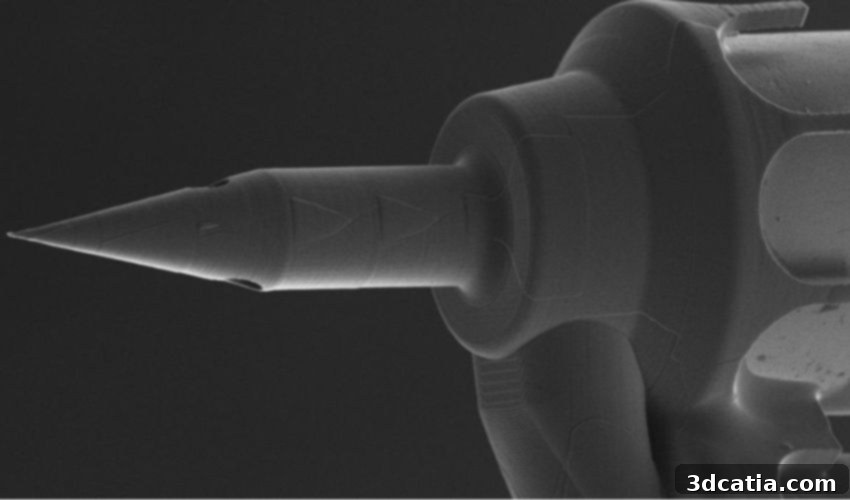
Revolutionizing Inner Ear Treatment: How 3D-Printed Microneedles Offer Precision for Hearing Loss and Cochlear Disorders
The inner ear, a marvel of biological engineering, plays a crucial role in both hearing and balance. Yet, its delicate and intricate anatomy, particularly the cochlea, has long presented one of medicine’s most formidable challenges. Its limited accessibility and complex structure create significant hurdles in effectively treating conditions such as hearing loss, hyperacusis, and various other inner ear disorders that profoundly impact millions worldwide. For over a decade, a pioneering team of physicians and engineers at Columbia University has dedicated itself to overcoming these inherent obstacles. Their persistent research has culminated in the development of a revolutionary microneedle, specifically engineered to dramatically enhance the precision and efficacy of medical treatments for hearing impairments. Central to the creation of this groundbreaking device has been the transformative power of additive manufacturing, a technology enabling unprecedented precision and efficiency in production.
This remarkable advancement is largely thanks to continuous innovations in 3D printing technologies. ENT surgeon Dr. Anil Lalwani, in collaboration with mechanical engineer Jeffrey Kysar, has successfully spearheaded the development of an ultra-thin, 3D-printed microneedle designed for highly localized, precision medicine within the inner ear. This innovation holds immense promise for improving current therapeutic approaches, particularly by enabling access to previously unreachable or highly sensitive areas of the cochlea. The microneedle’s exceptional design and capabilities were achieved through the application of two-photon photolithography, a specialized and cutting-edge 3D printing technique renowned for its ability to produce structures with incredibly high resolution and intricate detail. This sophisticated method ensures that the developed needle is significantly sharper and more refined than any existing medical instruments, while simultaneously maintaining the necessary robustness and structural integrity required for practical surgical application.
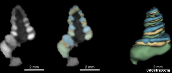
The microneedle can inject a contrast agent into the inner ear so that changes in the cochlea can be detected, helping to diagnose diseases such as Meniere’s disease. MRI images of the cochlea of a guinea pig show the different compartments of the cochlea. (Credit: Anil Lalwani/Columbia University Vagelos College of Physicians and Surgeons)
The Unparalleled Challenge of Cochlear Anatomy and Traditional Limitations
Understanding the true significance of this microneedle innovation requires delving into the profound difficulties presented by the cochlea’s unique anatomy. Dr. Lalwani eloquently describes it as a complex, spiral-shaped, fluid-filled bone – famously the hardest bone in the human body. This inherent robustness, paradoxically, makes it incredibly challenging to access and treat therapeutically. To reach damaged cells or introduce therapeutic agents, clinicians must navigate through an extraordinarily delicate membrane, often only about 2 millimeters wide. Traditional surgical instruments, designed for larger scales, frequently cause this fragile membrane to tear during insertion. Such damage is not merely an inconvenience; it can lead to severe complications, including further irreversible hearing loss or debilitating balance problems, thereby exacerbating the very conditions clinicians aim to treat. The research team recognized that to advance inner ear medicine, they first needed to profoundly understand the biomechanical factors contributing to this tearing. They conceptualized the membrane not as a simple barrier, but as a tightly stretched tarp, where the size and manner of piercing are critical. If the entry point created by an instrument is too large or blunt, the membrane will inevitably rupture. This crucial insight directly informed the design of their 3D-printed microneedle, whose final width is no wider than a human hair, ensuring minimal invasiveness and maximum precision.
Additive Manufacturing: The Foundation of Unprecedented Precision
The development of such a finely tuned instrument would be virtually impossible without the capabilities afforded by modern 3D printing, and specifically, the advanced technique of two-photon photolithography. This method represents a significant leap beyond conventional manufacturing, allowing for the fabrication of complex, three-dimensional structures with feature sizes down to the sub-micrometer scale. Unlike traditional methods that rely on subtractive processes or assembling pre-made parts, two-photon photolithography builds objects layer by layer by focusing femtosecond laser pulses into a photosensitive resin. This highly localized energy causes cross-linking only at the focal point, enabling the creation of intricate designs that are incredibly precise, sharp, and durable. For the inner ear microneedle, this meant the ability to design a device with a tip so fine it could pierce the delicate cochlear membrane without tearing it, yet robust enough to withstand the forces of insertion and fluid dynamics. The unprecedented control over geometry and material properties offered by this additive manufacturing process was indispensable in achieving the desired balance of sharpness, flexibility, and strength, ensuring the microneedle’s efficacy and safety for inner ear interventions. This innovative approach ensures that the barriers of micro-anatomy are no longer insurmountable, heralding a new era for otological procedures.
Broadening Therapeutic Horizons: Targeted Delivery and Diagnostic Breakthroughs
The primary objective behind the creation of this cutting-edge 3D-printed microneedle was to enable therapeutic interventions within the cochlea without incurring damage to its crucial membranes. This objective has been successfully met, opening doors to a new era of inner ear treatment. However, its utility extends far beyond mere access; the microneedle facilitates both advanced drug delivery and essential diagnostic procedures, promising a holistic improvement in patient care.
Targeted Drug Delivery for Inner Ear Disorders
One of the most significant applications of this innovative microneedle is its potential for highly targeted drug delivery. By allowing direct access to specific compartments within the cochlea, the device can deliver therapeutic agents precisely where they are needed most. This capability is paramount for treating a range of inner ear conditions, including various forms of hearing loss, tinnitus, and hyperacusis. Traditional systemic drug administration often means that only a small fraction of the medication reaches the inner ear, requiring higher doses that can lead to systemic side effects. With the microneedle, drugs – whether they are corticosteroids for inflammation, gene therapies for genetic hearing loss, or neuroprotective agents – can be delivered in smaller, more effective concentrations directly to the affected cells, minimizing adverse effects and maximizing therapeutic impact. This precision holds the key to personalized medicine for the inner ear, tailoring treatments to the exact needs of each patient at the cellular level. This direct approach not only enhances treatment efficacy but also significantly reduces the potential for systemic complications, marking a major step forward in therapeutic strategies.
Diagnostic Breakthroughs: Understanding Inner Ear Diseases
Beyond therapeutic delivery, the 3D-printed microneedle offers a critical advancement in diagnosing inner ear diseases. Its ability to safely and precisely remove fluid samples from the cochlea is revolutionary. Analyzing these fluid samples can provide invaluable biomarkers and insights into the pathological processes occurring within the inner ear, which are currently very difficult to obtain. For instance, this technique is particularly beneficial for diagnosing conditions like Meniere’s disease, a debilitating localized disorder characterized by a triad of symptoms: episodic vertigo (dizziness), tinnitus (ringing in the ear), and fluctuating sensorineural hearing loss, often accompanied by aural fullness. The exact cause of Meniere’s disease is still debated but is largely associated with an abnormal buildup of fluid (endolymph) in the inner ear. By enabling the precise aspiration and analysis of this fluid, clinicians can gain direct diagnostic evidence, potentially leading to earlier, more accurate diagnoses and the development of tailored treatment strategies. This capability moves us closer to understanding the underlying mechanisms of these complex conditions and offering more effective, individualized care, ultimately improving the quality of life for those affected.
Promising Results and Future Outlook
The journey from concept to clinical reality involves rigorous testing, and the 3D-printed microneedle has already demonstrated highly promising results in animal studies. Numerous operations have been successfully performed on animals without any discernible negative effects, such as further hearing loss or balance issues. Remarkably, the delicate cochlear membrane showed rapid healing, typically recovering within just two days after each procedure. This rapid healing and lack of adverse effects are strong indicators of the device’s safety and efficacy, paving the way for potential future human trials. The consistent success in these animal models underscores the microneedle’s potential to fundamentally transform inner ear treatment through truly precise surgical intervention. These preclinical results build a robust foundation for its eventual application in human patients, where the need for such precision is paramount.
Dr. Lalwani’s assertion that “It’s not an exaggeration to say our microneedle could be key to precision medicine for the inner ear” encapsulates the profound impact this innovation is expected to have. This technology represents a paradigm shift from broad, often ineffective, treatments to highly localized, targeted interventions. It promises to unlock new avenues for research into inner ear biology and pathology, leading to a deeper understanding of conditions that have long baffled medical science. As regulatory processes are navigated and human trials potentially commence, this 3D-printed microneedle stands poised to revolutionize otology, offering hope for millions suffering from challenging hearing and balance disorders. Its success also highlights the ever-growing importance of additive manufacturing and interdisciplinary collaboration between engineering and medicine in pushing the boundaries of what is medically possible. The future of inner ear health looks significantly brighter thanks to this pioneering work.
What are your thoughts on the groundbreaking 3D-printed microneedle for hearing loss treatment and other inner ear disorders? We invite you to share your perspectives in a comment below or join the conversation on our LinkedIn, Facebook, and Twitter pages! Don’t forget to sign up for our free weekly Newsletter here to receive the latest 3D printing news straight to your inbox! You can also find all our compelling videos and content on our YouTube channel.
*Cover Photo: Anil Lalwani/Columbia University Vagelos College of Physicians and Surgeons