
Revolutionizing Regenerative Medicine: Penn State Pioneers 3D Bioprinted Living Skin with Hair Follicles
The landscape of modern medicine is continually being reshaped by groundbreaking technological advancements, and among the most transformative is 3D printing. Its applications span an incredibly diverse range, from custom prosthetics and surgical guides to the ambitious frontier of bioprinted organs and tissues. In particular, the field of regenerative medicine has witnessed remarkable progress, offering new avenues for treating patients with complex injuries, burns, or congenital conditions that involve significant tissue loss. A recent and highly significant development comes from a dedicated research team at Penn State University. This pioneering team has successfully engineered and 3D bioprinted living skin that possesses the unprecedented potential to integrate functional hair follicles. This breakthrough utilized human tissue, specifically leveraging fat cells and their vital supporting structures to construct the sophisticated bio-ink necessary for the printing process. The immediate objective of this ambitious project was to demonstrate the efficacy and potential of this bioprinted skin by restoring injured rat skin to its original, healthy state, thereby laying crucial groundwork for future human applications and transforming the prospects for reconstructive surgery.
While the ability to produce rudimentary, thin layers of skin using 3D printing technology has been demonstrated in prior research, these earlier efforts often lacked the structural complexity and biological functionality required for true regenerative success. What sets the current Penn State study apart as a monumental leap forward is its unprecedented success in bioprinting a *complete and living system* comprising multiple, integrated skin layers. This includes the crucial lowest layer, known as the hypodermis. The hypodermis is a vital component of skin architecture, primarily composed of adipose (fat) tissue and supportive connective tissues. Beyond its well-known roles in insulation and energy storage, the hypodermis provides critical cushioning and structural support, particularly important for areas like the scalp and face, which are often prone to trauma and require robust underlying support. A truly innovative aspect of this advanced bioprinting technology is its capability for intraoperative application. This means the skin can potentially be printed directly onto an injury site during an ongoing surgical procedure, offering immediate, customized, and biologically integrated solutions. Interestingly, the researchers deliberately chose not to print the outermost visible skin layer, the epidermis. This strategic decision is based on a profound biological understanding: the epidermis possesses an inherent capacity for self-regeneration and naturally develops from the underlying dermal and hypodermal layers once the foundational structure is established. This strategic approach streamlines the printing process while capitalizing on the body’s intrinsic healing mechanisms, making the process more efficient and biologically harmonious.
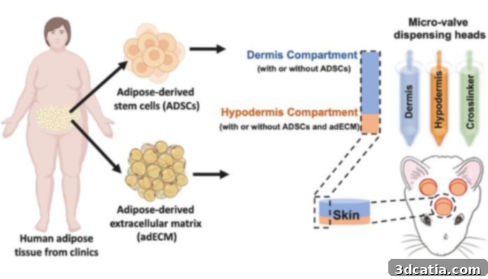
Photo Credits: Bioactive Materials
The groundbreaking results of this intricate research project were recently published in the esteemed scientific journal Bioactive Materials, garnering significant attention from the scientific community and beyond. Professor Ibrahim T. Ozbolat, a distinguished Professor of engineering science and mechanics, and of biomedical engineering, led this ambitious international research collaboration. He elaborated on the profound implications of their work, stating, “Reconstructive surgery to correct trauma to the face or head from injury or disease is usually imperfect, resulting in scarring or permanent hair loss. With this work, we demonstrate bioprinted, full thickness skin with the potential to grow hair in rats. That’s a step closer to being able to achieve more natural-looking and aesthetically pleasing head and face reconstruction in humans.” This statement underscores the critical need for improved reconstructive techniques and highlights how their bioprinting method addresses fundamental challenges that have long plagued patients undergoing such procedures. The ability to minimize scarring and restore natural hair growth represents a significant improvement in patient outcomes, not only in terms of physical healing but also psychological well-being, as appearance often plays a crucial role in a patient’s recovery and self-esteem.
How to Reconstruct Skin and Hair With 3D Bioprinting: A Detailed Look at the Methodology
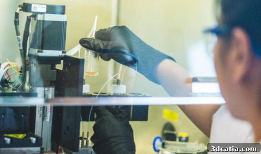
The innovative research project employed a sophisticated 3D bioprinter specifically designed with three distinct chambers, each meticulously filled with a different, critical biological component essential for tissue construction. The researchers began by sourcing adipose tissue, commonly known as fat tissue, from patients undergoing procedures at the Penn State Health Milton S. Hershey Medical Center. The primary aim was to carefully process this fatty tissue to extract its extracellular matrix (ECM). The ECM is a complex network of proteins and carbohydrates that provides structural and biochemical support to surrounding cells, giving tissues their shape, stability, and elasticity. This extracted ECM formed a fundamental part of the custom-designed bio-ink, acting as a natural scaffold for the new tissue. This crucial step highlights the bio-mimetic approach of the research, aiming to replicate the body’s natural cellular environment as closely as possible.
Furthermore, to produce the second indispensable component of the multi-faceted bio-ink, adult stem cells were also diligently extracted from the same fatty tissue. Adipose-derived stem cells (ADSCs) are highly valued in regenerative medicine due to their multipotent nature, meaning they possess the remarkable ability to differentiate into various cell types, including fat cells, cartilage, bone, and importantly for this study, cells relevant to skin regeneration and hair follicle formation. These stem cells are the living building blocks that would ultimately form the new tissue. The researchers filled the third pressure chamber with a carefully formulated coagulation solution. This solution plays a vital role in stabilizing the bioprinted structure immediately after deposition, allowing the bio-ink components to crosslink and form a stable tissue construct. Professor Ozbolat further elaborated on the precision and strategic advantage of this multi-chamber system:
“The three compartments allow us to co-print the matrix-fibrinogen mixture along with the stem cells with precise control. We printed directly into the injury site with the target of forming the hypodermis, which helps with wound healing, hair follicle generation, temperature regulation and more.”
As Professor Ozbolat emphasized, the hypodermis is not merely a passive layer but is directly involved in several vital physiological processes. Crucially, it facilitates the intricate process by which stem cells mature and differentiate into functional fat cells, a process indispensable for robust wound healing. Beyond wound repair, the hypodermis plays a pivotal, active role in the complex hair follicle cycle, particularly in initiating and promoting new hair growth. This understanding guided the team’s approach. In their meticulous research, Ozbolat and his team discovered that the coordinated and precise co-supply of both the extracellular matrix and the stem cells is absolutely critical for the successful formation and maturation of a functional hypodermis. This synergistic delivery system ensures that the nascent tissue receives all the necessary biological cues and structural support to develop correctly, demonstrating a sophisticated mastery over tissue engineering principles.
Furthermore, the detailed experiments conducted revealed compelling insights into the cellular interactions at play. It was observed that the presence and activity of the fat cells within the bioprinted construct likely altered the surrounding extracellular matrix in a specific, beneficial manner. This modification is believed to have rendered the microenvironment significantly more supportive and conducive to the successful formation and integration of hair root structures. These profound findings from the Penn State researchers thus hold immense potential, promising a transformative impact on the field of reconstructive facial surgery, particularly for patients suffering from severe trauma, burns, or oncological resections that result in significant tissue loss. Moreover, this technology offers a beacon of hope for developing effective treatments for hair growth restoration in humans within injured or diseased areas, addressing a long-standing challenge in dermatology and plastic surgery. The ability to regenerate a complete, hair-bearing skin unit could dramatically improve both the functional and aesthetic outcomes for countless individuals. For those interested in delving deeper into the scientific intricacies of this pioneering work, further details can be found in the published study HERE.
The implications of this breakthrough in 3D bioprinting extend far beyond the immediate applications. This research pushes the boundaries of tissue engineering, demonstrating the feasibility of creating complex, multi-layered living tissues with specialized functions. It opens new avenues for studying skin diseases, developing more accurate drug testing models, and ultimately, moving closer to the ambitious goal of bioprinting entire organs for transplantation. While challenges remain, such as scaling up production, ensuring long-term graft stability, and navigating stringent regulatory approvals, the Penn State team’s work represents a monumental step forward. This innovative approach to skin regeneration, combining advanced 3D printing with cutting-edge biological engineering, signifies a promising future where personalized, regenerative solutions become a standard of care for complex medical conditions, offering unprecedented hope for patients facing significant tissue damage and loss. The integration of hair follicles into the bioprinted skin adds another layer of complexity and functionality, moving regenerative medicine closer to restoring not just structure, but also natural aesthetics and protective features.
What do you think of this incredible advancement in 3D printed skin with the potential for functional hair follicles? We invite you to share your thoughts and perspectives in a comment below or connect with us on our LinkedIn, Facebook, and Twitter pages! Don’t forget to sign up for our free weekly newsletter here to receive the latest 3D printing news straight to your inbox! You can also find all our engaging videos and interviews on our YouTube channel, exploring the dynamic world of additive manufacturing.
*Cover Photo Credits: Penn State