
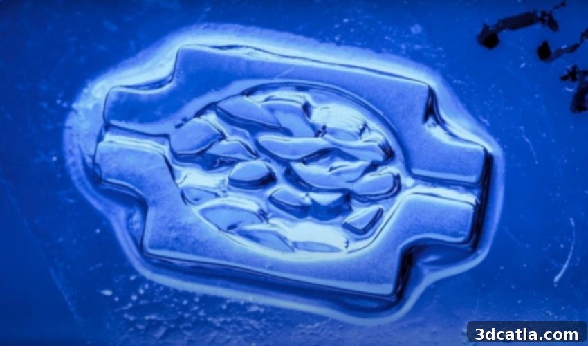
Revolutionizing Regenerative Medicine: UC San Diego Pioneers High-Resolution DLP 3D Bioprinting for Vascularized Human Tissues
The future of medicine is rapidly being reshaped by groundbreaking advancements in additive manufacturing, particularly within the realm of 3D bioprinting. A distinguished research team from the Jacobs School of Engineering at the University of California, San Diego, is at the forefront of this revolution, meticulously developing a novel method of 3D bioprinting. This innovative approach is fundamentally based on digital light processing (DLP) technology, a technique renowned for its precision and speed. The ambitious project’s core objective is to engineer sophisticated vascularized 3D tissues that, despite being meticulously fabricated in a laboratory setting, can mimic the intricate functionality of human-like living cells, complete with complex biomaterial structures. This experimental study, a testament to relentless scientific inquiry, was recently published in the esteemed journal *Science Advances*. The publication offers an in-depth explanation of how these dedicated researchers successfully unveiled one of the most promising avenues in 3D bioprinting, specifically focusing on the use of advanced biocompatible materials to achieve unprecedented results.
DLP 3D printing has, for some time, stood out as one of the most widely adopted and impactful technologies within the medical sector. Its implementation across various medical domains has showcased immense potential for a broad spectrum of biomedical applications. These critical applications span vital areas such as accelerated drug development and screening, the creation of viable organs for transplantation, and the advancement of both regenerative medicine and highly personalized therapeutic approaches. However, despite its remarkable capabilities, this branch of the health sector has historically contended with significant practical and technical limitations inherent to conventional additive manufacturing processes. These challenges have included, but were not limited to, the daunting task of printing tissues with exceptionally finely resolved structures while simultaneously maintaining high cell densities – a critical requirement for functional tissue mimics. The ability to precisely replicate the delicate architecture of natural human tissues, particularly the intricate vascular networks essential for nutrient and oxygen transport, has remained a particularly formidable hurdle. Recognizing these inherent difficulties, the pioneering team at the Jacobs School opted for a distinctly different, highly innovative approach, seeking to overcome these long-standing obstacles.
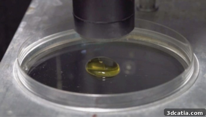
By carefully reducing bio-ink density, the problematic effect of light scattering is effectively avoided, leading to higher resolution prints (Credits: UC San Diego / David Baillot)
Advancing Regenerative Medicine: The Creation of Vascularized Human Tissues
The nanoengineering team at UC San Diego has achieved a monumental breakthrough, successfully realizing a sophisticated 3D bioprinting system capable of fabricating vascularized human tissues with unprecedented fidelity. This cutting-edge, patented technology is ingeniously founded on a process of additive layering, meticulously building biological structures and complex tissues using a precise combination of living cells and advanced biopolymers. A persistent challenge in bioprinting has been the relationship between bio-ink density and print resolution. Typically, a higher density of bio-ink, while desirable for cell concentration, leads to increased light scattering during the DLP process. This scattering effect significantly hinders the resolution of the printed structure, making it difficult to achieve the fine details required for functional tissues, especially those with intricate vascular networks. To address this fundamental limitation, the researchers developed and utilized a specially formulated, highly biocompatible polymer tailored for high-resolution DLP 3D printing. The true innovation, however, lies in their ability to dramatically reduce the light scattering effect by a factor of ten, thanks to the strategic inclusion of ‘iodixanol’ – a novel contrast agent integrated directly into the bio-ink. This crucial advancement enabled them to print constructs with exceptionally high cell densities while simultaneously achieving remarkably high resolution, a combination previously considered a major technical hurdle in the field.
The bioprinting process itself involves a carefully orchestrated and complex series of repeated steps, culminating in the precise accumulation of numerous layers that collectively form the desired 3D model. A key component of this intricate methodology is the meticulous adjustment of the refractive index of the bio-ink. By precisely tuning this property, the researchers were able to minimize the detrimental light scattering effect, thereby significantly enhancing the overall manufacturing process and print quality. Through this sophisticated control, the team successfully achieved an impressive resolution of 50 micrometers (µm) using a bio-ink formulation primarily based on gelatin methacrylate (GelMA). GelMA is a versatile biomaterial, often favored in tissue engineering for its excellent biocompatibility and tunable mechanical properties. Crucially, the refractive index of their optimized GelMA-based bio-ink was precisely matched to accommodate an exceptionally high cell density, reaching up to 0.1 billion cells per milliliter. This capability is paramount for creating truly functional tissues. Professor Shaochen Chen, the distinguished leader of this groundbreaking project, eloquently explains the subsequent critical phase: “After printing, we culture the construct to allow the cells to mature or reorganize into a functional tissue. Therefore, the cell is like a seed, and each cell type has a specific density at which they are most potent to sprout.” This analogy beautifully illustrates the biological principles at play, emphasizing that the initial printing creates a scaffold of ‘seeds,’ which then need the right environment and time to ‘sprout’ and develop into viable, functional tissue structures. The precision of the printing, combined with the ideal cell environment, allows for the self-organization and maturation that is essential for complex biological function.
The far-reaching implications of this innovative research are profound and promise to significantly accelerate and strengthen various facets of modern medicine. Firstly, it could revolutionize drug development by enabling the creation of more accurate and physiologically relevant *in vitro* tissue models. These models would provide a superior platform for screening new drug candidates, leading to more effective therapies and potentially reducing the need for animal testing. Secondly, this technology offers a beacon of hope for alleviating the critical shortage of organ donors worldwide. By bioprinting personalized, functional organs, the lengthy and often fatal waitlists for transplants could be drastically reduced. Furthermore, the ability to use a patient’s own cells for bioprinted organs could effectively eliminate the pervasive challenge of immune rejection, a major complication in current organ transplantation procedures.
Looking ahead to the not-too-distant future, this pioneering technology holds immense promise for enabling the widespread development of sophisticated *in vitro* tissue models characterized by remarkably high cell density and functional vasculature. These advanced models will not only serve as invaluable tools for fundamental biological research but also pave the way for a new era of personalized medicine, where treatments can be tailored with unprecedented precision to individual patient needs. The ability to create functional tissue units could ultimately lead to novel regenerative therapies for repairing damaged tissues and organs, offering hope for patients suffering from chronic diseases or injuries. While these transformative applications are still progressing through research and development phases, the foundation laid by the UC San Diego team marks a significant leap forward. In the meantime, those interested in delving deeper into the scientific intricacies of this remarkable achievement can access the full study, as published in *Science Advances*, by clicking HERE.
What are your thoughts on this groundbreaking new method for creating vascularized human tissues? Do you foresee this technology rapidly changing the landscape of organ transplantation and regenerative medicine? We encourage you to share your insights and opinions in a comment below or join the conversation on our LinkedIn, Facebook, and Twitter pages! For the very latest updates, news, and analyses from the world of additive manufacturing delivered directly to your inbox, don’t forget to sign up for our free weekly Newsletter here. You can also explore all our engaging videos and in-depth content on our official YouTube channel, where we continually bring you the most exciting developments in 3D printing technology.
*Cover photo credits: UC San Diego / David Baillot