
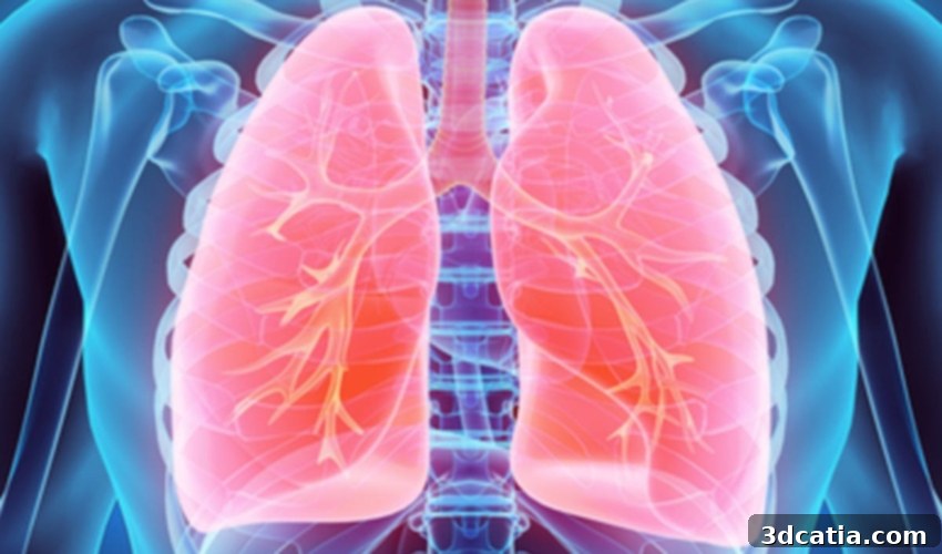
Pioneering Respiratory Research: POSTECH Unveils Advanced 3D Bioprinted Lung Model for Disease Studies
As the gentle transition from winter to spring unfolds, the blossoming flowers and the return of warmer weather often bring with them an unwelcome guest: dust. While largely imperceptible to most, this seasonal dust can be a significant source of distress and an exacerbating factor for individuals suffering from various respiratory diseases. The past few years, profoundly marked by the global COVID-19 pandemic, have cast an even longer shadow over those with vulnerable respiratory systems, amplifying concerns about airborne pathogens and environmental irritants. The human lung, a marvel of biological engineering, possesses an intricate and delicate structure, characterized by its extreme thinness and complex alveolar network. This inherent complexity, crucial for efficient gas exchange, simultaneously poses immense challenges for scientific research, particularly when it comes to developing accurate and experimentally viable artificial lung models. Such models are an indispensable tool for understanding disease mechanisms, testing new therapeutic agents, and ultimately, finding cures for conditions affecting these fragile and vital organs.
Recognizing this critical need, a collaborative team of distinguished researchers from the Pohang University of Science & Technology (POSTECH) has achieved a groundbreaking milestone in medical science. Professor Sungjune Jung from the Department of Materials Science and Engineering, alongside Professor Joo-Yeon Yoo and Ph.D. candidate Dayoon Kang from the Department of Life Sciences, have successfully engineered a sophisticated three-dimensional lung model. This innovative model, which incorporates a diverse array of human alveolar cell lines, was meticulously created using state-of-the-art inkjet bioprinting technology. Professor Jung elaborated on the significance of their achievement, stating, “While our team has a track record of printing cells and fabricating tissues using bioprinting methods, this represents a world-first in simulating a complex alveolar barrier with a three-layer structure, meticulously crafted to approximately 10 μm thickness.” He further emphasized the unprecedented nature of their work by adding, “Crucially, this is also the inaugural instance where an artificial alveolar barrier has been successfully infected with a virus, allowing for the direct observation of a physiological antiviral response within a controlled laboratory setting.” This capability opens up unparalleled opportunities for studying viral pathogenesis and evaluating antiviral treatments with unprecedented accuracy.
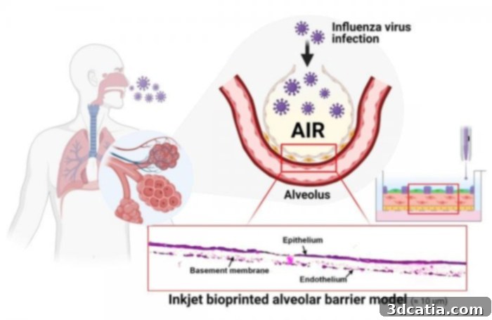
An intricate inkjet bioprinted alveolar barrier model, representing a significant leap in organ-on-a-chip technology. (Photo Credit: POSTECH)
The Precision and Potential of Inkjet Bioprinting for Medical Research
The choice of inkjet bioprinting technology was pivotal to the POSTECH team’s success, primarily due to its inherent advantages in precision, scalability, and customization. This advanced bioprinting method offers a unique capacity to produce not only standardized tissue models for broad research applications but also patient-customized tissues, tailored to individual biological characteristics. Furthermore, its potential for mass production signifies a paradigm shift, enabling the widespread availability of these sophisticated models. These capabilities position inkjet bioprinting as a transformative force, poised to potentially replace conventional, often less accurate and more labor-intensive methods of manufacturing test models, such as 2D cell cultures or animal testing. The research team specifically leveraged a technique known as ‘drop-on-demand’ inkjet bioprinting. This method allowed for the remarkably high-resolution deposition of alveolar cells, enabling them to construct the complex three-layer alveolar barrier model with an astounding thickness of approximately 10 micrometers (μm). This level of precision is critical for accurately mimicking the native tissue environment within the human lung.
What truly sets this novel alveolar barrier model apart is its remarkable ability to faithfully reproduce physiological responses that occur at the actual tissue level. This includes critical aspects such as viral infectivity and the subsequent antiviral response mounted by the cells. To demonstrate its efficacy and potential, the researchers successfully utilized the bioprinted model as an influenza virus infection platform. Through this experimental setup, they were able to directly observe key biological phenomena, including the self-proliferation of the virus within the artificial tissue and the host cells’ intrinsic antiviral responses. This capability is invaluable for virological studies, allowing scientists to gain deeper insights into how respiratory viruses interact with lung tissue and how the body responds at a cellular and molecular level. The fidelity of these observed responses underscores the model’s potential as a powerful tool for foundational research into respiratory pathogens and host-pathogen interactions.
Unlocking New Avenues for Respiratory Disease Research and Drug Development
The implications of POSTECH’s 3D bioprinted lung model extend far beyond foundational research. As Professor Jung articulated, “The artificial tissue we have successfully produced can serve as an invaluable early-stage platform for robustly evaluating the efficacy of therapeutic drugs and vaccines designed to combat infectious respiratory viruses. This includes critically important pathogens like the COVID-19 virus, as well as seasonal influenza and other emerging viral threats.” The ability to enable mass production of these models, combined with stringent quality control mechanisms, ensures that a reliable and consistent supply of experimental platforms can be maintained. This is a crucial factor for large-scale drug screening and vaccine testing initiatives, which often require thousands of identical models. Furthermore, the capacity to fabricate patient-customized disease models is a game-changer for the burgeoning field of personalized medicine. By using cells derived from specific patients, researchers can create models that accurately reflect an individual’s unique physiological and pathological characteristics. This allows for the testing of drugs and therapies in a personalized context, potentially leading to more effective treatments and fewer adverse reactions.
This innovation is particularly timely, given the ongoing global challenges posed by respiratory pandemics. The traditional lengthy and often cost-prohibitive process of drug discovery and vaccine development can be significantly accelerated by having such accurate and scalable in vitro models. These models can help identify promising compounds earlier, screen out ineffective ones more quickly, and reduce reliance on animal testing, which often fails to fully recapitulate human physiological responses. Beyond drug efficacy, these models also offer unprecedented opportunities for understanding the fundamental mechanisms of viral infection, progression, and resolution within human lung tissue. Researchers can meticulously study how viruses replicate, how they damage cells, and how the immune system responds, all within a highly controlled and reproducible environment. This detailed understanding is essential for developing targeted therapies and preventative strategies against a wide range of respiratory ailments.
Synergies in 3D Printing for Medical Applications: Beyond Bioprinting
While POSTECH’s achievement focuses on functional, living tissue models through bioprinting, it’s important to acknowledge the broader impact of 3D printing technologies across various medical applications. In a related yet distinct endeavor, another company called Axial3D recently undertook a project that utilized Formlabs’ SLA (Stereolithography) 3D printing technology to create highly accurate anatomical replicas. Their work involved replicating the lungs of a COVID-19 patient, but not for functional testing. Instead, these models served as crucial visual and tactile aids for medical professionals, allowing them to better understand the extent and nature of the damage caused by the virus to a patient’s lungs. Such physical models are invaluable for pre-surgical planning, educating patients and their families, and for training medical students. They provide a tangible representation of complex internal structures, which can enhance diagnostic accuracy and improve surgical outcomes. This highlights the versatile utility of 3D printing in medicine, ranging from the creation of functional biological tissues to highly detailed anatomical models, each serving different but equally vital purposes in advancing healthcare.
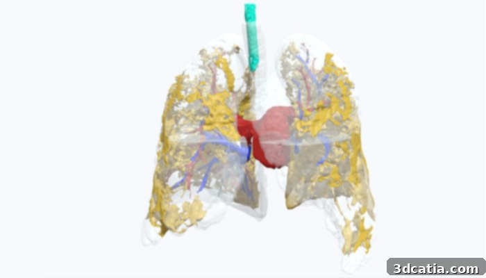
Axial3D’s precision model of a COVID-19 patient’s lung, utilized for enhanced medical visualization and planning. (Photo Credit: Axial3D)
The Future of In Vitro Disease Modeling and Personalized Medicine
The development of POSTECH’s 3D bioprinted lung model represents a significant stride towards the future of medical research and patient care. This innovation is a powerful testament to the potential of tissue engineering and bioprinting to bridge the gap between traditional research methods and the complex realities of human physiology. As these technologies continue to evolve, we can anticipate even more sophisticated and integrated organ-on-a-chip systems that can mimic multiple interacting organs, offering a holistic view of systemic diseases. The ability to create accurate, functional, and patient-specific disease models promises to dramatically accelerate the pace of scientific discovery, reduce the ethical and practical limitations associated with animal testing, and ultimately lead to faster development of life-saving treatments and vaccines. The promise of personalized medicine, where treatments are precisely tailored to an individual’s genetic makeup and disease characteristics, becomes much more tangible with such advanced modeling platforms. This research not only offers a new tool for combating respiratory viruses but also paves the way for a deeper understanding of lung biology and pathology, potentially transforming how we approach a myriad of respiratory conditions, from asthma and COPD to idiopathic pulmonary fibrosis and lung cancer. The meticulous design and successful validation of this alveolar barrier model underscore the innovative spirit driving modern biomedical engineering, offering a breath of fresh air for those striving to improve global health outcomes.
For those interested in delving deeper into the specifics of POSTECH’s groundbreaking 3D printed lung model and the intricate details of their research methodology, the full press release is available HERE. This comprehensive document provides additional scientific context and insights into the team’s innovative approach.
What are your thoughts on this remarkable 3D bioprinted lung model and its potential impact on medical research and patient care? We invite you to share your perspectives and engage in the conversation by leaving a comment below. You can also connect with us and share your opinions on our social media platforms: Facebook, Twitter, and LinkedIn pages! Stay informed about the latest advancements and breaking news in the exciting world of 3D printing by signing up for our free weekly Newsletter here, delivered directly to your inbox!
*Cover photo courtesy of: MDGRPHCS