Applications
HUCA Team Uses 3D Printing to Treat Aortic Aneurysms
According to the Spanish Society of Cardiovascular and Endovascular Surgery, a significant portion of the population over 60 is at high risk of developing an abdominal aortic aneurysm. When these lesions affect areas with branches leading to vital organs, treating…
- 3 min read
- Applications
- Six more stories
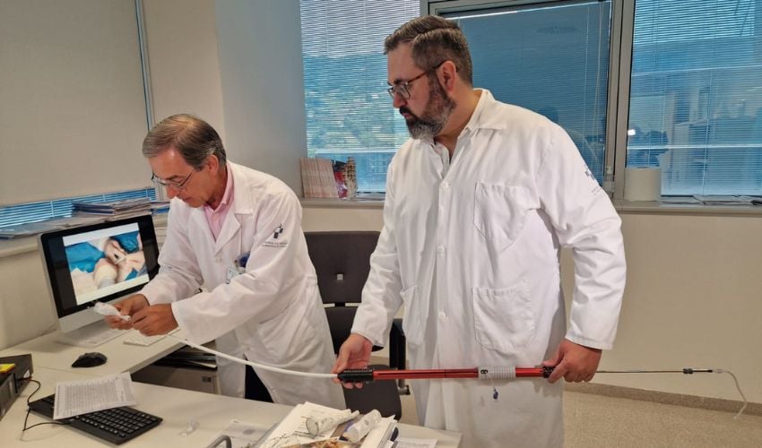
According to the Spanish Society of Cardiovascular and Endovascular Surgery, a significant portion of the population over 60 is at high risk of developing an abdominal aortic aneurysm. When these lesions affect areas with branches leading to vital organs, treating them becomes a real puzzle for surgeons. In Oviedo, the Central University Hospital of Asturias (HUCA) has found a solution: using 3D printing to create customized prostheses and implanting them through a minimally invasive procedure.
An aneurysm is, in simple terms, a bulge that forms in an artery. In the case of the abdominal aorta, the large artery that carries blood from the heart to the rest of the body, the risk is high. The challenge increases when the aneurysm is located near the branches supplying blood to the liver, kidneys, or intestines. In these cases, treatment requires maintaining blood flow to these organs while simultaneously reinforcing the damaged artery. Open surgery is an option, but it involves large incisions, long hospital stays, and high risk for older patients or those with other health conditions. The less invasive alternative, endovascular treatment, uses prostheses inserted through the arteries, but they don’t always fit well in complex anatomies.
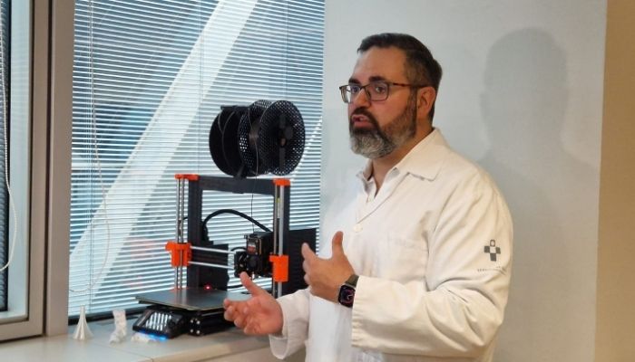
Dr. Francisco Álvarez Marcos, Associate in the Angiology and Vascular Surgery Department at the Central University Hospital of Asturias (HUCA)
To overcome these limitations, a team from the Angiology and Vascular Surgery Department at HUCA, led by Dr. Manuel Alonso, turned to 3D printing. Using a scan, they accurately reproduced a patient’s aorta in a physical model, following the “normal” blood flow for surgical planning. On this model, the medical team modifies a conventional prosthesis, creating openings exactly where the visceral arteries connect. Before entering the operating room, the design is checked using a paper template that mimics the prosthesis structure. This extra step reduces errors and ensures a perfect fit. The major advantage is achieving a level of customization similar to conventional prostheses, but in much less time and precisely tailored to the patient’s anatomy.
More from the desk
Six other notes from the rest of the bench.
 NewsA canteen 3D food printer in a primary schoolIn the UK, Big Bang Fair researchers have implemented a canteen 3D food printer. The 3D printer creates a variety of dishes for students. The goal is to encourage them to pursue a career in STEM (Science, Technology, Engineering and Mathematics). Teaching 3D…
 News3D Systems to Return to the Extrusion Market Thanks to Two New AcquisitionsOnly two months into the year and the additive manufacturing industry continues to show its vitality and growth. Already, we have seen launches and more from leading companies in the sector and we now have a new one to add…
 NewsTop 5 Videos of the Week: the 3D printing trends for 2020 and much more!This week we found the top 5 videos about 3D printing and the way additive manufacturing is shaping our society. The technology has infiltrated many sectors, from medical to automotive! Check out our selection of the week to keep up with the innovations! This week we…
 ApplicationsThe University of Sydney Takes a Leap Towards Functional 3D Printed Human TissuesEmbarking on a journey that seems ripped from the pages of science fiction, a recent breakthrough has brought us one step closer to the future of 3D printing functional human tissues. A collaborative effort between bioengineers and biomedical scientists from…
 GuidesWho shaped the additive manufacturing market in 2019?The year 2019 is coming to an end, with good results for the additive manufacturing market. This market is becoming increasingly industrialized because the technology is becoming a production-ready process, less reliant on rapid prototyping activity like it was 3…
 PrintersWhy Combine 3D Printing and Industrial Automation Applications?3D printing is widely used in the field of automation such as in compression molding. It’s a formative manufacturing method, currently popular in industry, that involves compressing the desired amount of molding material between two heated presses to give it…