Applications
A 3D Printed Hip Prosthesis Has Been Successfully Implanted in Poland
Additive manufacturing is increasingly known for its many benefits in medicine. Time and again, it has made it possible to produce perfectly fitted parts for every person, a necessity especially when it comes to parts like prostheses. Now, this has…
- 3 min read
- Applications
- Six more stories
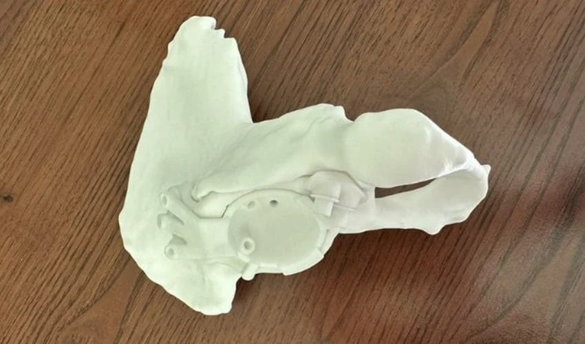
Additive manufacturing is increasingly known for its many benefits in medicine. Time and again, it has made it possible to produce perfectly fitted parts for every person, a necessity especially when it comes to parts like prostheses. Now, this has been demonstrated once again at a hospital in Lublin, a city in Poland, where 3D printing was successfully used for the first time to make a hip prosthesis.
After a patient experienced complications with her previously inserted steel prosthesis, the medical team needed to find a quick solution. “As a result of the loosening of the earlier placed prosthesis of the hip joint, in this particular female patient, there came to be a large defect in the pelvic bones,” explained Dr. Andrzej Atras, head of the hospital’s traumatology-orthopedics department. According to Dr. Atras, such an operation is very difficult to handle. There must be an exact distribution of forces in the bone socket so as not to cause degeneration of the bone tissue in the future, but rather to promote reconstruction and regeneration of the bone bed. “We had to use a specially expanded implant which would allow for stable embedding in the bone with the use of the remaining regions which contained good quality fragments of bone,” he continued. A 3D printed hip prosthesis was the solution.
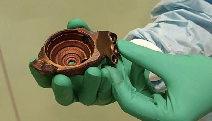
The original steel prosthesis loosened over time (photo credits: Dziennik Wschodni)
3D Printing the Hip Prosthesis
With the help of the orthopedic company Medgal, the team developed a 3D printed implant for the hip joint that was perfectly adapted to the patient’s needs. First, an X-ray and high-resolution computerized tomography were taken, after which the 3D model of the pelvis and hip joint could be created. This allowed doctors to assess the defects and damage in the patient’s pelvic floor and, with the help of a team of engineers and an orthopedic surgeon, make the implant. This was eventually fitted to the remaining acetabulum, allowing the acetabular component of the prosthesis to be placed firmly in the pelvic bone. In an interview, Dr. Atras noted, “We worked on that socket for two months, to give it the correct geometry and biocompatibility with the patient’s body. It often required several hour long consultations and an evaluation of many variants of the construction in order to select the most optimal one.”
More from the desk
Six other notes from the rest of the bench.
 Printers3D Printing: The Future of ConstructionIn 2004, Professor Behrokh Khoshnevis of the University of South Carolina attempted the first 3D printed wall. Since then, this innovation has exploded and it’s now possible to build a house in just 20 hours! The professor developed an FDM…
 ApplicationsNew Study Examines if EU Regulations Are Stifling Medical 3D PrintingThe medical 3D printing market has grown sharply in recent years. This includes in the EU, where the overall healthcare AM market is expected to grow at a CAGR of 15.4% until 2029. Unsurprising when you consider that the European…
 NewsTop 5 Videos: Ford Motor Company Large-Format 3D PrintingIn this week’s compilation of 3D printing videos, discover how the Ford Motor Company is using large-format 3D printing, resulting in the company saving over 90% manufacturing time! Next, you will go behind the scenes of Mighty Buildings, an American…
 NewsTop 5 Videos: 3D Printing With CoffeeAs the sun sets on another week of September living, we gather together a selection of handpicked videos for your viewing pleasure. Like a digital vineyard, we roam the hills and valleys of the internet to bring you the most…
 ApplicationsApplication of the Month: Lamborghini Uses 3D printing for the SC63 Hypercar at the 24 Hours of Le Mans 2024The use of 3D printing in motor racing has become increasingly common. The ability to rapidly produce, customize and select the most appropriate materials for each application has led many automakers to adopt this technology. In Formula 1, for example,…
 Printers#3DStartup: Rosotics and the World’s Largest Metal 3D PrinterUS-based startup Rosotics specializes in the development of 3D equipment, with a particular focus on metal additive manufacturing. Its goal? To offer manufacturers the largest metal 3D printer ever imagined on today’s market. Called the M1, this manufacturing platform offer