News
Study Suggests Benefits of 3D Bioprinting In Plant Cell Research
A recent study from North Carolina State University experimented using 3D bioprinting to produce plant cells in order to assess the use of the method in biological research. They produced Arabidopsis- (thale cress; often used in genetic research) and soybean…
- 3 min read
- News
- Six more stories
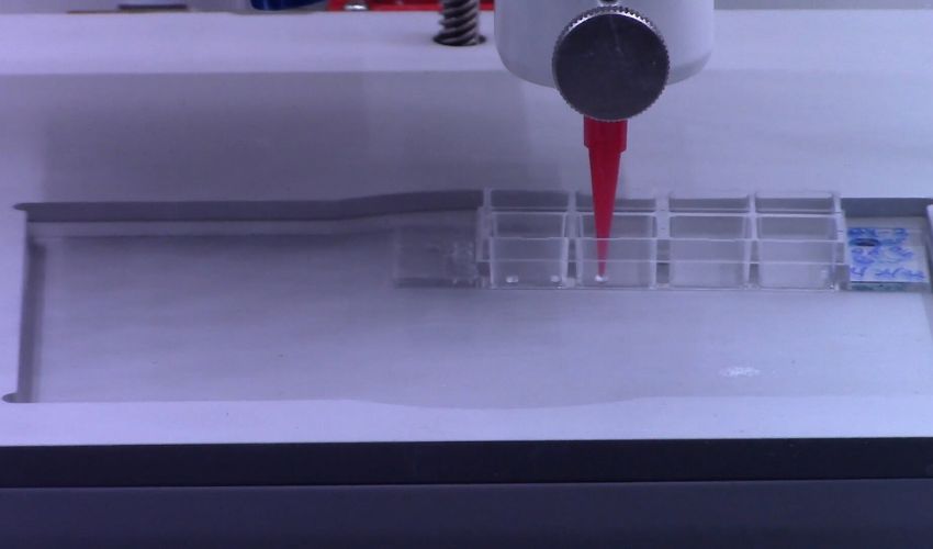
A recent study from North Carolina State University experimented using 3D bioprinting to produce plant cells in order to assess the use of the method in biological research. They produced Arabidopsis- (thale cress; often used in genetic research) and soybean cells. Their 3D-printed cells had adequate viability and went on to produce microcalli (groups of plant tissue), indicating that they were successful productions. These findings suggest that 3D bioprinting could be at least as effective as traditional methods for plant cell reproduction.
Methods
Firstly, let’s look at the method used to conduct the experiment: pneumatic extrusion. This is a type of 3D bioprinting in which the researcher utilizes air pressure to force out the biological material through a nozzle. This printed both the bioink and the support structures, likened to scaffolding. For the latter, they tested two different types- agarose and sodium alginate, to assess which one proved more effective. The soy bean cells showed good viability of almost 49% two weeks after creation, which indicates that bioprinting could produce cells with equal viability to traditional manual pipetting. After 5 days, viability within the 3D bioprinted structure was almost 25% for the Arabidopsis cells with agarose ‘scaffolding’.
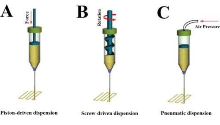
The team used a bioprinting method called pneumatic extrusion or dispension – C. Image credit: Advanced Nanobiomed Research on ResearchGate
For the viable cells, researchers were able to assess production of microcalli (groups of unorganized parenchyma cells, a type of plant cell). The cell cycle reentry coincided with the induction of core cell cycle genes and genes related to cell regeneration. Essentially, they caused the bioprinted cells to restart their cell cycles and reproduce. They found that after 14 days, on average 90% of bioprinted constructs formed 5-6 microcalli. This indicates that 3D bioprinted cells have the potential to undergo cell division and behave as cells produced by traditional methods.
More from the desk
Six other notes from the rest of the bench.
 PrintersZOZOSUIT, the Suit with an Integrated 3D Body Scanner For Weight TrackingZOZOFIT is a fitness technology brand and Japan’s largest fashion e-commerce company, recognized worldwide for its use of new technologies in its products. Its latest innovation is the world’s first affordable 3D body scanning technology, the suit they have named&hell
 News3D Printed Wheel System Makes Life Easier for Shelly the TurtleStudents of the third to sixth grade at Prairie Queen Elementary School in Papillion, USA, conducted a community science project that used additive manufacturing to help a physically challenged turtle. The students, who are all members of the school’s 3D…
 NewsPolySLS project achieves 67% energy savingsINEOS Styrolution has released findings from an R&D project, named PolySLS, which is focused on developing a new energy saving styrenics based material for additive manufacturing. INEOS Styrolution is a world leading styrenics supplier, produced for a wide range of…
 MaterialsGeneral Lattice Awarded Contract to Improve Combat Helmet Impact Absorption for United States ArmyCombat helmets are one of the oldest pieces of battle armor, however, this vital piece of personal equipment is long overdue for re-design and improvement. The United States military has been harnessing the power of additive manufacturing to innovate and…
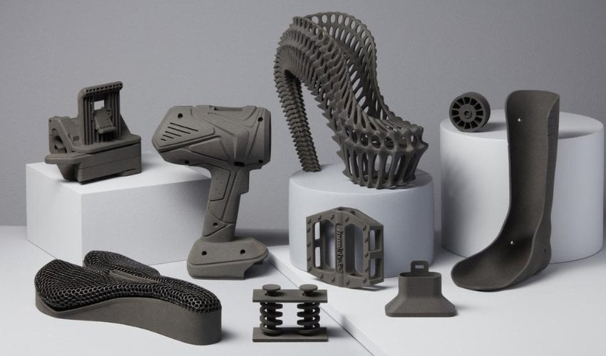 NewsEverything You Should Know About Rapid Prototyping in 3D PrintingThe product development landscape is constantly evolving, demanding innovative approaches to bridge the gap between ideas and reality. Rapid prototyping (RP) has emerged as a powerful tool in this realm, fostering creativity and efficiency. At its core, rapid prototyping is&helli
 NewsMcDonald’s will offer completely 3D printed burgers by 2020Fast food giant McDonald’s has just announced the creation of a brand new 3D printed burger, its 3D Mic Mac. Restaurants should be equipped with 3D printers by the end of 2020 in order to offer this customisable hamburger. According…