
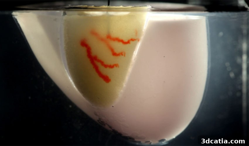
Revolutionizing Regenerative Medicine: Harvard’s SWIFT Technique for Bioprinting Vascularized Organs
The ambitious field of regenerative medicine is constantly striving to overcome significant biological hurdles, particularly in the quest to engineer functional human organs. A groundbreaking development from a team of researchers at Harvard University’s Wyss Institute for Biologically Inspired Engineering and the John A. Paulson School of Engineering and Applied Sciences (SEAS) is bringing us closer to this reality. They have unveiled a novel solution that enables the bioprinting of intricate blood vessels directly onto living tissues, addressing a critical challenge that has long hampered progress in organ engineering. Their innovative technique, known as SWIFT (Sacrificial Writing Into Functional Tissue), was detailed in their seminal paper titled ‘Biomanufacturing of organ-specific tissues with high cellular density and embedded vascular channels,’ published in the prestigious journal Science Advances. This pioneering approach holds immense promise for the creation of larger, more complex organs that can effectively deliver vital oxygen and nutrients, moving beyond the limitations of current lab-grown tissue constructs.
For many decades, scientists globally have dedicated their efforts to cultivating human organs in laboratory settings. Despite significant advancements, a pervasive challenge has been the inability to grow organs that are not only adequately sized for human transplantation but also possess the sophisticated internal structures, like vascular networks, essential for effective oxygen and nutrient transport. Without a robust blood supply, cells in larger tissue constructs quickly die due to starvation and waste accumulation. While the journey is still in its nascent stages, remarkable progress is being made. Not long ago, a team of researchers in Israel garnered international attention when they successfully 3D printed a small human heart, complete with cells, blood vessels, and chambers, from a patient’s own biological materials. This monumental achievement underscored the immense potential of bioprinting technologies to transform the landscape of organ transplantation and regenerative therapies. While such advancements undoubtedly make bioprinting a tremendously promising avenue for creating replacement human organs, it is crucial to acknowledge that the field is still at the very beginning of realizing its full therapeutic impact.
The urgency for these innovative solutions cannot be overstated. The global organ transplant crisis continues to inflict immense suffering and loss of life. In the United States alone, a stark reality unfolds daily: an average of 20 individuals tragically die each day while awaiting an organ transplant. Despite the impressive efforts that see more than 30,000 organ transplants performed annually, the number of patients on organ waitlists continues to climb, currently exceeding 113,000. This growing disparity between the demand for transplantable organs and their scarce availability highlights a profound medical and ethical challenge. The development of functional, lab-grown organs could dramatically reduce these waiting lists, offering a new lease on life to countless patients and alleviating the immense burden on healthcare systems. Harvard’s SWIFT technique represents a significant leap forward in addressing this critical unmet need, offering a pathway to engineer tissues that are not only viable but also capable of integrating seamlessly into the human body.
The Revolutionary SWIFT Technique: Building Functional Tissues from the Inside Out
The SWIFT technique, meticulously developed by the brilliant minds at Harvard’s Wyss Institute and SEAS, represents a paradigm shift in bioprinting. This innovative method focuses on precisely 3D printing vascular channels – the microscopic conduits that mimic natural blood vessels – directly into living tissue matrices. These matrices are ingeniously constructed from stem-cell-derived Organ Building Blocks (OBBs), which are essentially miniature, self-assembling aggregates of cells organized to recapitulate the architecture and function of specific organ tissues. This approach cleverly circumvents the traditional difficulty of bioprinting entire organs cell by cell. Mark Skylar-Scott, one of the co-first authors of the study, eloquently articulated the core philosophy behind SWIFT: “Rather than trying to 3D print an entire organ’s worth of cells, SWIFT focuses on only printing the vessels necessary to support a living tissue construct that contains large quantities of OBBs, which may ultimately be used therapeutically to repair and replace human organs with lab-grown versions containing patients’ own cells.” This strategic focus on vascularization – the lifeblood of any organ – is what distinguishes SWIFT and unlocks its potential for creating truly functional and viable tissue constructs for therapeutic applications.
At its heart, the SWIFT technique is an elegant two-step process, each stage meticulously designed to overcome critical challenges in tissue engineering. The foundational first step involves the formation of a dense, living matrix composed of these specialized Organ Building Blocks. These OBBs are carefully aggregated to create a tissue construct that closely mimics the high cellular density characteristic of natural human organs. Sébastien Uzel, another co-first author, elaborated on the dual benefit of this initial phase: “Forming a dense matrix from these OBBs kills two birds with one stone: not only does it achieve a high cellular density akin to that of human organs, but the matrix’s viscosity also enables printing of a pervasive network of perfusable channels within it to mimic the blood vessels that support human organs.” This high cellular density is paramount because it ensures that the engineered tissue has the cellular complexity and functional capacity required for physiological relevance. Furthermore, the inherent viscosity of this dense OBB matrix provides the ideal medium for the subsequent sacrificial writing process, allowing for the precise creation of intricate vascular networks.
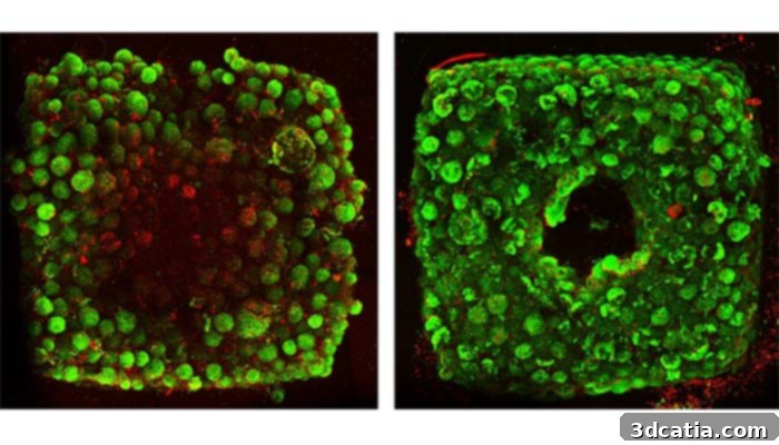
Organ-specific tissues that were printed with embedded vascular channels using SWIFT remained viable (right), while tissues grown without these channels experienced cell death within 12 hours (left).
The second, equally crucial step of the SWIFT technique leverages the principle of sacrificial 3D printing. This stage relies on maintaining the dense OBB matrix at carefully controlled, cold temperatures. At these lower temperatures, the matrix possesses an ideal consistency: it is sufficiently soft and pliable to be manipulated without causing damage to the delicate cells within, yet robust enough to firmly hold its structural shape. This unique rheological property makes it the perfect medium for the subsequent printing process. A fine nozzle precisely navigates through this cold, viscous matrix, depositing a specialized strand of gelatin-like ink. As this ink is extruded, it gently pushes the living cells aside, creating a temporary pathway without inflicting any cellular harm. The magic happens next: once the sacrificial ink has been printed to form the desired vascular network, the entire cold matrix is carefully warmed to physiological temperature, approximately 37°C. At this temperature, the OBB matrix begins to stiffen, consolidating its structure and becoming more solid-like, thus permanently locking the newly formed channels in place. Simultaneously, the gelatin-based ink, which was specifically chosen for its temperature-dependent properties, melts away. This molten gelatin can then be easily washed out, leaving behind a perfectly formed, interconnected network of open channels embedded seamlessly within the tissue construct. These bioprinted channels are remarkably versatile, with diameters that can be precisely controlled, ranging from a mere 400 micrometers to a full 1 millimeter, mimicking the diverse sizes of capillaries and larger vessels found in the human body. The paramount importance of these channels lies in their ability to be continuously perfused with oxygenated media and essential nutrients, thereby sustaining the life and function of the cells deep within the engineered tissue.
The efficacy of the SWIFT technique was rigorously tested by the Harvard scientists, yielding highly encouraging results that underscore its transformative potential. During their experiments, they observed a critical difference: organ-specific tissues that were successfully printed with these intricate, embedded vascular channels using the SWIFT method remained remarkably viable and functional over extended periods. In stark contrast, control tissues that were grown without the benefit of these essential vascular channels suffered widespread cell death within a mere 12 hours. This direct comparison dramatically demonstrated the absolute necessity of an integrated vascular network for sustaining engineered tissue constructs of clinically relevant size and complexity. One of the most compelling tests involved assessing whether the bioprinted tissues could exhibit organ-specific functions, which is the ultimate goal of regenerative medicine. The research team meticulously printed, evacuated, and perfused a network of channels into a matrix meticulously crafted from heart-derived cells. They then continuously flowed nutrient-rich media through these channels for over a week. The results were astounding: “During that time, the cardiac OBBs fused together to form a more solid cardiac tissue whose contractions became more synchronous and over 20 times stronger, mimicking key features of a human heart.” This remarkable display of enhanced contractile strength and synchronized beating is a monumental step towards developing functional cardiac patches or even larger heart tissue replacements, offering hope for patients suffering from cardiovascular diseases.
This research indeed marks a very promising stride in the field of bioprinting and tissue engineering. The ability to create functional, vascularized tissues opens up new frontiers for drug testing, disease modeling, and ultimately, therapeutic applications in humans. The full scientific details and methodologies can be explored in the comprehensive paper, accessible HERE. This represents not just an incremental improvement but a foundational advancement that could pave the way for a future where organ transplantation challenges are significantly alleviated, and personalized regenerative therapies become a reality. The team’s ongoing work will undoubtedly focus on scaling up these constructs, ensuring long-term viability, and refining the integration of these engineered tissues into living systems, bringing us closer to a future free from organ waiting lists.
We’d love to hear your thoughts on the revolutionary SWIFT technique and its implications for the future of medicine! Feel free to share your insights in a comment below, or join the conversation on our Facebook and Twitter pages. To stay informed about all the latest breakthroughs in 3D printing and bioprinting, don’t forget to sign up for our free weekly Newsletter, delivering cutting-edge news directly to your inbox!