
Pioneering Internal Bioprinting: How a Miniature Robotic Arm is Transforming Surgical Tissue Repair
Globally, millions of individuals annually face the debilitating effects of tissue damage, stemming from a wide array of diseases, traumatic injuries, or unforeseen medical conditions. This pervasive issue places immense strain on healthcare systems and significantly impacts patients’ quality of life. For decades, the primary method for addressing such internal damage during surgical procedures has relied heavily on traditional techniques like suturing (stitches) or stapling to approximate and promote tissue healing. While these methods have been foundational in surgical practice, they are not without their limitations. The delicate nature of internal organs, the potential for sutures to fail, the risk of complications such as infection, inflammation, scarring, and prolonged recovery times often present significant challenges, sometimes leading to re-operations or long-term health issues. Recognizing these critical unmet needs in modern medicine, a team of visionary researchers at the University of New South Wales (UNSW) in Australia has embarked on a groundbreaking endeavor. They have successfully developed a miniature, highly flexible robotic arm designed to revolutionize internal surgery by directly repairing damaged human body tissue or organs through advanced 3D bioprinting techniques. This innovative approach promises to offer unprecedented precision and efficacy, ushering in a new era for regenerative medicine and minimally invasive surgical interventions.
The advent of 3D printing, and more specifically 3D bioprinting, has rapidly emerged as one of the most transformative technologies in the medical sector over the past decade. Its applications are continuously expanding, impacting everything from cutting-edge research to complex surgical planning and even direct therapeutic interventions. The field of 3D bioprinting, in particular, holds immense promise for the future of regenerative medicine. Numerous ambitious projects worldwide are actively leveraging this technology to replicate intricate cellular structures, engineer functional tissues, and even create complex organs of the human body for transplantation, drug testing, and disease modeling. This capability offers a unique avenue for understanding complex biological processes, developing personalized treatments, and potentially eliminating organ donor shortages. Beyond direct human applications, the versatility of 3D bioprinting extends into diverse industries, showcasing its broad applicability. A compelling example comes from the cosmetics industry, where the renowned beauty company Chanel has innovatively utilized this technology. Chanel developed 3D bioprinted skin models not for aesthetic enhancement in the traditional sense, but to meticulously investigate various skin conditions, rigorously test the efficacy and safety of new ingredients, and ultimately improve its extensive product line with greater scientific precision and ethical considerations, avoiding animal testing. This diverse range of applications underscores the profound impact and multifaceted potential of 3D bioprinting, setting the stage for innovations like the UNSW robotic arm.
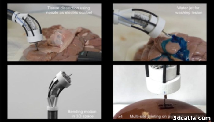
The F3DB 3D bioprinter boasts several innovative features for internal tissue repair. (Image credits: University of New South Wales)
The F3DB: A Breakthrough in Miniature Robotic 3D Bioprinting
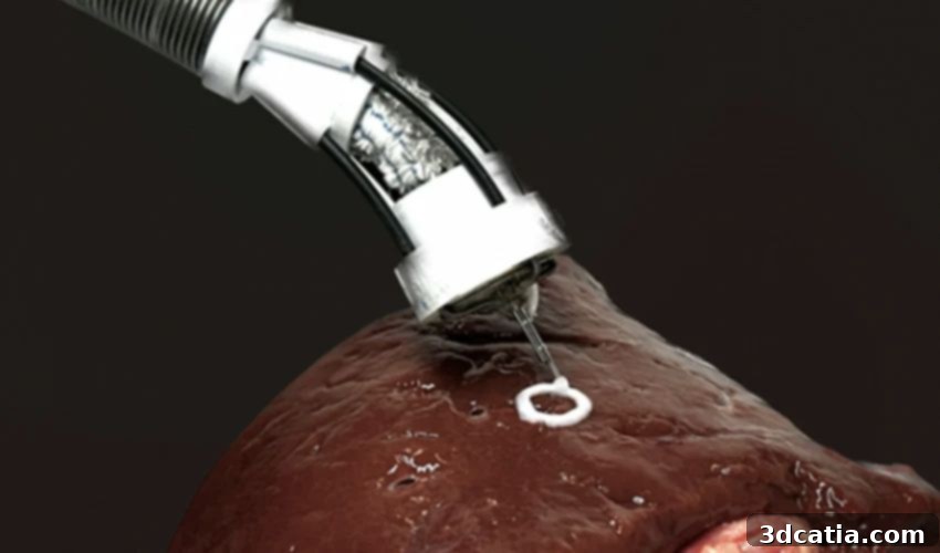
At the heart of this transformative medical innovation lies the device known as the F3DB – a moniker that encapsulates its function as a Flexible 3D Bioprinter. This remarkable miniature bioprinter is the brainchild of a dedicated team, spearheaded by the brilliant minds of Dr. Thanh Nho Do and Dr. Mai Thanh Thai from UNSW. Their vision was to create a surgical tool capable of unprecedented precision and accessibility within the human body. The F3DB is ingeniously designed to be remarkably tiny and incredibly flexible, enabling its insertion into the body through existing natural openings or via minimal, small incisions, thereby drastically reducing the invasiveness of surgical procedures. Once positioned, this sophisticated device can meticulously apply specialized bio-materials directly onto the surface of damaged internal organs and tissues. The core of the F3DB’s advanced capability is its state-of-the-art print head. This component features a three-axis rotating mechanism, granting it exceptional dexterity and control within the confined spaces of the human anatomy. This precision print head is securely attached to the end of a remotely controllable robotic arm, allowing surgeons or medical professionals to manipulate its movements with exquisite accuracy from outside the body. The nozzle of the F3DB is not only highly versatile but also incredibly intelligent; it can be programmed to print pre-designed shapes and patterns with high fidelity, ensuring consistent and reproducible tissue repair. Furthermore, for situations demanding nuanced control or adapting to unforeseen anatomical variations, the device can also be manually manipulated by the operator, offering a crucial layer of flexibility and adaptability for more complex surgical operations. This dual capability of programmed automation and manual override ensures optimal performance across a spectrum of surgical scenarios.
Unlocking Access and Precision: The F3DB’s Therapeutic Potential
One of the most significant advancements offered by the F3DB is its unparalleled ability to navigate and operate within anatomical regions previously deemed inaccessible or extremely challenging for conventional surgical instruments. The UNSW researchers highlight that the F3DB can effectively reach and perform repairs in critical internal organs such as the colon, stomach, heart, and bladder – areas where existing rigid or less flexible surgical devices often fall short, limiting therapeutic options and necessitating more invasive approaches. The F3DB’s innovative design, characterized by its miniature dimensions, is crucial to this expanded accessibility. The smallest prototype currently developed measures approximately 11-13 mm, placing it squarely within the size range of traditional endoscopes. This compact form factor allows for insertion through natural orifices or minimal incisions, significantly reducing patient trauma and accelerating recovery. However, the ambitious research team is not resting on its laurels; they are actively committed to developing even smaller iterations of the F3DB for future medical applications, pushing the boundaries of minimally invasive surgery even further. Beyond its primary role in tissue repair, the F3DB demonstrates remarkable potential in advanced therapeutic procedures, including the removal of certain cancers. Specifically, the researchers envision its use in procedures like endoscopic submucosal dissection (ESD) for conditions such as colon cancer. ESD is a sophisticated technique employed by gastroenterologists to meticulously remove early-stage cancerous lesions or precancerous growths from the gastrointestinal tract, preserving the integrity of the organ. In this intricate process, the F3DB would enable highly localized and precise targeting. The technique often involves using a water jet to create a fluid cushion beneath the lesion, lifting it from the muscular layer and facilitating its clean, complete removal. Integrating the F3DB’s bioprinting capabilities could potentially enhance this process by not only facilitating lesion removal but also immediately printing regenerative biomaterials onto the resected surface, promoting faster healing and reducing the risk of recurrence or complications. This dual functionality marks a monumental leap in the management of internal pathologies, offering a future where complex interventions are less invasive and more effective.
The Path Forward: Testing, Enhancements, and Clinical Adoption
The development journey of the F3DB has progressed through crucial validation stages, demonstrating its foundational viability and promise. Initial testing conducted by the UNSW researchers involved deploying the device within an artificial colon model, meticulously simulating the internal environment and challenges of a human gastrointestinal tract. Concurrently, a diverse range of biomaterials, selected for their biocompatibility and regenerative properties, were applied and printed onto the surface of a pig’s kidney, providing a realistic ex vivo model for assessing direct tissue interaction. Crucially, these preliminary tests yielded highly encouraging results: the printing process, despite its precision and localized application, did not exhibit any negative impact on the surrounding cells, confirming the F3DB’s potential for safe and effective bio-integration. Building on this success, the immediate next phase of research involves the rigorous testing of the F3DB in live animal models. This vital step will provide invaluable insights into its performance in a dynamic, complex biological system, evaluating factors like long-term tissue regeneration, inflammatory responses, and overall safety profile in vivo. Furthermore, the engineering team is dedicated to enhancing the F3DB’s capabilities by integrating several critical features. Plans include embedding a high-resolution camera directly into the device, offering surgeons real-time visual feedback from within the body, which is essential for accurate navigation and precise bioprinting. Even more advanced is the envisioned system for reconstructing high-fidelity images of *moving* tissue inside the body. This feature is paramount for organs like the heart or those affected by respiration and peristalsis, allowing the bioprinter to dynamically adapt and print with unwavering precision despite physiological movements. The aspirations for this technology are ambitious yet grounded in scientific progress: within the next five to seven years, the innovative engineering team at the University of New South Wales hopes to see the F3DB adopted by medical professionals worldwide. This widespread adoption would fundamentally revolutionize the way internal surgery is performed, moving towards less invasive, more regenerative, and significantly more effective patient treatments. The full scientific study detailing the intricacies of this project and its findings is available for deeper exploration HERE. For a more accessible, synthesized overview of the project as published by the Australian University, interested readers can click HERE.
Join the Conversation on the Future of Medical Bioprinting
The development of the F3DB by UNSW researchers represents a monumental stride forward in medical technology and regenerative medicine. By offering a solution for precise, minimally invasive internal tissue repair and therapeutic interventions, this miniature robotic 3D bioprinter has the potential to redefine surgical standards, significantly improve patient outcomes, and usher in an era where complex internal damage is addressed with unprecedented precision and efficacy. This innovation stands as a testament to the power of combining advanced robotics with cutting-edge bioprinting to solve some of healthcare’s most persistent challenges.
What are your thoughts on this groundbreaking bioprinting project and its implications for the future of surgery? We invite you to share your insights and engage with our community. Leave a comment below or connect with us on our social media channels: LinkedIn, Facebook, and Twitter. Don’t miss out on the latest advancements and news in the world of 3D printing; sign up for our free weekly Newsletter here to get updates delivered straight to your inbox. You can also explore all our informative videos and engaging content on our YouTube channel.
*Cover photo credits: University of New South Wales