
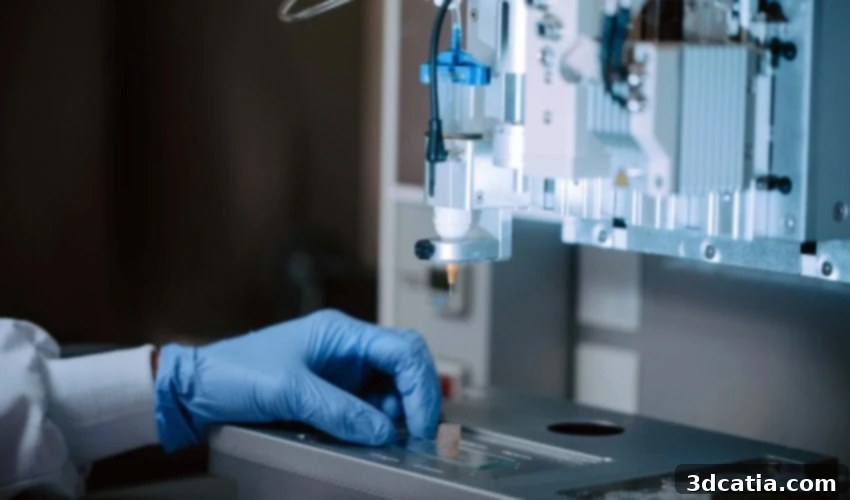
3D Bioprinting Revolutionizes Medical Research with Advanced Human Tissue Models
The ethical debates surrounding animal experimentation in scientific research have persisted for decades, with valid concerns raised about the welfare of the animals involved. In Spain alone, an alarming figure of approximately 1,144,214 animals is utilized annually for scientific purposes, highlighting the sheer scale of this practice. While animal models have historically been indispensable tools in medicine for understanding diseases and testing potential treatments, they inherently possess significant limitations. The fundamental biological differences between species mean that animal physiology doesn’t always accurately replicate human anatomy and disease progression, leading to results that may not translate effectively to human patients. This scientific inadequacy, coupled with the ethical imperative to minimize animal suffering, drives the urgent need for viable alternatives.
Enter 3D printing – a transformative technology emerging as a powerful solution with the potential to dramatically reduce, and eventually replace, animal experimentation across various scientific disciplines. Its application has already demonstrated promising results in sectors like cosmetics and pharmaceuticals, where more accurate and ethical testing methods are paramount. A groundbreaking recent development comes from the research center CIC biomaGUNE in the Basque Country. Their innovative study delves into the creation of highly realistic tissue models, complete with artificial blood vessels, utilizing advanced 3D bioprinting techniques combined with cutting-edge nanomaterials. This pioneering work represents a significant leap forward in tissue engineering and offers a compelling pathway towards more humane and effective medical research.
Leading the Way in Bioprinting Innovation and Animal Experimentation Alternatives
At the forefront of this remarkable scientific endeavor are Dr. Dorleta Jimenez de Aberasturi, Dr. Uxue Aizarna, and Dr. Malou Henriksen. These visionary researchers, integral members of the Biofunctional Hybrid Materials group at the prestigious CIC biomaGUNE center, have dedicated their expertise to pushing the boundaries of tissue engineering. Their extensive work involves meticulously testing and refining a diverse array of techniques aimed at designing and fabricating increasingly realistic tissue models – models that can accurately serve the intricate demands of contemporary scientific research. The ultimate goal is to create platforms that can reliably predict human responses to drugs, understand disease progression, and pave the way for regenerative medicine, all while significantly reducing the reliance on animal testing.
A particular focus of their groundbreaking research has been the development of tissues incorporating sophisticated artificial blood vessels. The human vascular system is incredibly complex, with arteries and veins forming a critical network for nutrient and oxygen delivery, waste removal, and communication throughout the body. Mimicking this complexity in vitro is a monumental challenge. However, the team at CIC biomaGUNE has achieved this by creating artificial vessels that not only replicate the intricate structural architecture of human arteries but also exhibit dynamic functionality, such as the ability to pulse in response to external stimuli. This level of biomimicry is crucial because the mechanical forces exerted by blood flow play a vital role in cellular behavior and tissue development. Achieving such realistic models opens up unprecedented avenues for drug discovery, disease modeling, and even the future of organ transplantation, offering a more ethically sound and scientifically relevant approach than traditional animal models.
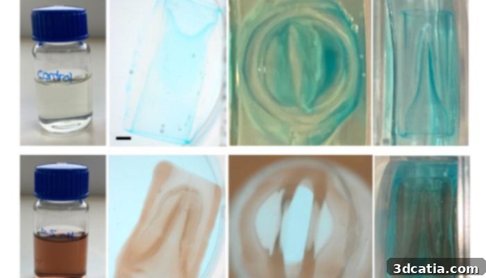
Valves manufactured through volumetric 3D printing. (Credit: American Chemical Society)
The Ingenious Bio-Ink: Merging Biology and Nanotechnology for Advanced Bioprinting
A cornerstone of this revolutionary advancement lies in the sophisticated composition of the “bio-ink” – the printable material that forms the very foundation of these artificial tissues. Unlike conventional inks, bio-inks must be precisely engineered to meet stringent biological and mechanical requirements, ensuring compatibility with living cells and the ability to form stable, functional structures. The CIC biomaGUNE team ingeniously chose methacrylated gelatin as their base material. Gelatin, derived from collagen, is highly favored in bioprinting due to its excellent biocompatibility, biodegradability, and natural extracellular matrix-like properties, which are crucial for cell adhesion, proliferation, and differentiation within a 3D construct. By chemically modifying gelatin through methacrylation, its mechanical properties can be precisely tuned, and it can be efficiently cross-linked (hardened) using light, allowing for the stable formation of complex 3D structures that mimic native tissues.
What truly sets this bio-ink apart is the strategic incorporation of gold nanoparticles. These minuscule particles, typically ranging from 1 to 100 nanometers, are known for their unique physical and chemical properties, making them invaluable in advanced biomaterials. In this application, gold nanoparticles play a pivotal role in enabling the enhanced functionalities of the printed tissues. They not only enhance the mechanical stability of the printed constructs, making them more robust and resilient under physiological conditions, but also contribute to the electrical conductivity of the material. This conductivity is significant for mimicking biological signals within tissues, particularly in nerve or muscle cells. Crucially, the presence of gold nanoparticles allows for unparalleled control over the behavior of the printed structure post-fabrication. This control can involve modulating cellular responses, guiding tissue maturation, or enabling dynamic responsiveness to external stimuli, effectively bridging the gap between inert scaffolding and living, functional tissue. This innovative blend of a cell-friendly hydrogel with responsive nanomaterials creates a versatile and highly advanced platform for engineering intricate and functional biological constructs with potential for wide-ranging medical applications.
Advanced Bioprinting Techniques: Building Functional Complexity for Drug Discovery
Beyond the groundbreaking bio-ink, the researchers at CIC biomaGUNE, in strategic collaboration with leading academic institutions, have pioneered two distinct and equally impressive bioprinting techniques that are critical to the functionality of these advanced tissue models. These innovations collectively allow for the creation of intricate biological structures that were previously unattainable with conventional methods, significantly contributing to the field of drug discovery and personalized medicine.
1. “Arteries in the Air”: Precision with Embedded Bioprinting for Vascularization
The first remarkable innovation is what the team refers to as “arteries in the air,” developed in close collaboration with Maastricht University. This technique tackles one of the most significant challenges in bioprinting: working with soft, delicate biomaterials. Many ideal biological materials for tissue engineering are hydrogels that lack sufficient mechanical integrity to hold complex shapes, especially when printing intricate, hollow structures like blood vessels. Traditional bioprinting methods often struggle with gravitational collapse or deformation when attempting to print such soft materials in free space, making it difficult to achieve the precise geometries required for functional vascularization.
To overcome this, the researchers employed an embedded bioprinting technique. In this method, the bio-ink is extruded directly into a supportive, sacrificial material – typically a viscous gel – rather than into open air. This surrounding gel provides immediate mechanical support, preventing the delicate “arteries” from collapsing during the printing process. The rheological properties of the support bath are carefully matched to allow for precise deposition of the bio-ink while simultaneously holding the printed structure in place. Once the printing is complete and the bio-ink is cross-linked (solidified) through a photopolymerization process, the sacrificial gel can be carefully removed, often by temperature change or chemical dissolution, leaving behind a perfectly formed, freestanding vascular structure. This approach allowed the team to precisely construct vessel models featuring the complex concentric layers characteristic of a real artery, including the distinct tunica intima, media, and adventitia, which are essential for physiological function, nutrient transport, and accurate biomimicry in drug testing platforms.
2. High-Speed Functional Valves with Volumetric 3D Printing for Dynamic Tissue Models
The second pivotal technological advancement involves the development of high-speed, functional valves, achieved through a collaboration with Utrecht University, utilizing cutting-edge volumetric 3D printing. Traditional 3D printing methods, including many bioprinting techniques, build objects layer by excruciating layer. While effective for many applications, this layer-by-layer approach can be slow and may introduce anisotropic properties or surface roughness that are undesirable in delicate biological constructs, especially those requiring smooth internal lumens for fluid flow.
This innovative volumetric technique operates by projecting the entire 3D shape of an object into a volume of photo-curable bio-ink simultaneously. Instead of slowly building up slices, light rays are precisely modulated and directed from multiple angles to harden the entire volume of the desired structure at once, akin to “sculpting with light.” This significantly accelerates the printing process from minutes or hours to mere seconds, making it ideally suited for creating sensitive biological constructs containing live cells, as prolonged exposure to stress can compromise cell viability. More importantly, this volumetric approach enables the creation of complex geometries with superior resolution, intricate internal features, and remarkably smooth surfaces, both internally and externally. This capability was instrumental in designing and integrating sophisticated valves into the tissue models – valves that are not merely static but are engineered to actively open and close in response to specific external stimuli, such as changes in pressure or electrical signals. The ability to create such dynamic, responsive components is a monumental step towards developing truly functional artificial organs and advanced “organ-on-a-chip” systems, offering unparalleled platforms for research, personalized medicine, and therapeutic applications without the need for animal subjects.

Part of the Biofunctional Hybrid Materials group at the Basque center. (Credit: CIC biomaGUNE)
Paving the Way for a New Era in Biomedical Science and Ethical Research
Summarizing their groundbreaking achievements, the researchers aptly state, “By combining these improved materials and various printing techniques, we managed to produce more complex structures that are much more similar to those found in the human body.” This declaration underscores the significant progress made in bridging the gap between simplified in vitro models and the intricate reality of human biology. The ability to create functional, biomimetic tissues with integrated vascular networks and dynamic valves marks a pivotal moment in tissue engineering and regenerative medicine, heralding a future with less reliance on animal models.
While these results undoubtedly represent a monumental scientific advance, the team, with characteristic scientific prudence, cautions that the journey is far from over. The process of engineering living, functional tissues is extraordinarily complex, encompassing challenges ranging from ensuring long-term cellular viability and efficient nutrient supply within the constructs to achieving scalability for widespread use and navigating the rigorous pathways of regulatory approval. Each incremental improvement in bio-ink formulation, printing resolution, or functional integration presents its own set of formidable engineering and biological challenges. Yet, the persistent dedication of researchers like those at CIC biomaGUNE continues to push the boundaries of what is possible in the quest for more ethical and effective scientific tools.
Despite the remaining hurdles, the implications of this research are truly enormous and far-reaching, promising to revolutionize several critical areas of biomedical science:
- Drug Discovery and Development: More accurate human tissue models can serve as superior platforms for screening new drug compounds, predicting their efficacy and toxicity with greater precision than traditional animal models or simplified cell cultures. This could significantly reduce the time and cost associated with bringing new medicines to market, while simultaneously improving patient safety and reducing animal harm.
- Disease Modeling: By replicating specific human tissues and their pathologies, these advanced models can offer unprecedented insights into the mechanisms of diseases like cardiovascular conditions, cancer, and diabetes. Researchers can study disease progression, test therapeutic interventions, and unravel complex biological pathways in a controlled, human-relevant environment, leading to more targeted therapies.
- Personalized Medicine: In the future, it may be possible to create patient-specific tissue models using a patient’s own cells. This revolutionary approach would enable clinicians to test various treatments directly on the patient’s “mini-organ” or tissue construct before administering them, leading to truly personalized, highly effective therapeutic strategies with minimal side effects.
- Regenerative Medicine and Organ Transplantation: The ultimate vision for bioprinting includes the creation of fully functional organs for transplantation, effectively addressing the critical shortage of donor organs. While this remains a long-term goal, the development of functional vascular networks and dynamic components like valves is a fundamental and essential step toward engineering whole, viable organs that can be safely integrated into the human body.
- Reducing Animal Experimentation: Perhaps most immediately impactful, these human-relevant models offer a direct and ethical alternative to animal testing, aligning with global efforts to adopt the 3Rs (Replace, Reduce, Refine) in research, thereby fostering a more humane and scientifically robust approach to biomedical discovery.
This pioneering research from CIC biomaGUNE, in collaboration with Maastricht and Utrecht Universities, represents a beacon of innovation, propelling us closer to a future where medical breakthroughs are achieved with greater precision, ethics, and human relevance. For those keen to delve deeper into the scientific intricacies of this work, the original scientific article can be explored HERE.
What are your thoughts on this incredible breakthrough in 3D bioprinting and its potential to transform medical research? Share your insights and comments below, or connect with us on our LinkedIn or Facebook pages. Don’t miss out on the latest advancements in additive manufacturing – subscribe to our free weekly Newsletter to receive top 3D printing news directly in your inbox. For more visual content, explore our extensive library of videos on our YouTube channel.
Cover Photo Credit: CIC biomaGUNE