
Revolutionizing Plant Science: The Power of 3D Bioprinting for Cellular Research
The field of plant biology is constantly seeking innovative methods to understand fundamental cellular processes, tissue development, and responses to environmental stimuli. Traditional techniques, while foundational, often present limitations in terms of precision, scalability, and the ability to create complex, controlled three-dimensional environments for cellular studies. Against this backdrop, a groundbreaking study from North Carolina State University has illuminated the immense potential of 3D bioprinting as a transformative tool for plant cell research. This pioneering experiment successfully utilized advanced bioprinting techniques to produce viable plant cells, specifically from Arabidopsis thaliana (commonly known as thale cress, a model organism in genetic research) and soybean, demonstrating a significant leap forward in our capacity to manipulate and study plant tissues in unprecedented ways. The successful production of viable cells that subsequently formed microcalli—groups of nascent plant tissue—suggests that 3D bioprinting could not only match but potentially surpass the effectiveness of conventional methods for plant cell propagation and analysis, opening new avenues for scientific discovery in agriculture and biotechnology.
The Innovative Methodology: Pneumatic Extrusion Bioprinting
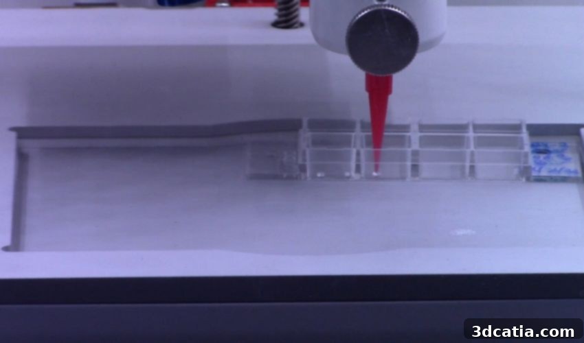
At the heart of this remarkable study was the application of pneumatic extrusion, a sophisticated form of 3D bioprinting. This technique leverages precisely controlled air pressure to meticulously dispense biological material, known as bioink, through a fine nozzle. This allows for the precise deposition of cells and their accompanying support structures, effectively building a 3D cellular construct layer by layer. The process is akin to using a scaffold to guide the growth and organization of the biological material, providing a structural framework that is critical for maintaining cell viability and facilitating proper tissue formation.
Bioink and Scaffold Design for Optimal Cell Growth
A crucial element in successful bioprinting is the careful selection of both the bioink—which contains the living cells—and the support structures, often referred to as ‘scaffolding.’ These scaffolds are vital for providing mechanical stability, mimicking the extracellular matrix found in natural tissues, and allowing for nutrient diffusion. In their experiments, the researchers rigorously tested two distinct types of hydrogel materials for these support structures: agarose and sodium alginate. Agarose, a polysaccharide derived from seaweed, is widely used in biological research due to its inertness and ability to form stable gels. Sodium alginate, another seaweed-derived polysaccharide, is favored for its biocompatibility and ability to gel rapidly in the presence of calcium ions, making it highly versatile for bioprinting applications. By comparing these two materials, the team aimed to determine which offered superior conditions for cell survival and proliferation within the printed constructs.
The results highlighted the efficacy of this approach. Soybean cells exhibited impressive viability, with nearly 49% remaining alive two weeks post-bioprinting. This viability rate is particularly significant as it demonstrates that 3D bioprinting can achieve results comparable to traditional, more labor-intensive manual pipetting methods, while offering greater control and potential for automation. For Arabidopsis cells, which are often more delicate, viability within the 3D bioprinted structure reached almost 25% after five days when agarose was used as the scaffolding material. These viability figures are not just encouraging; they signify a robust foundation for further research and application of 3D bioprinting in plant biology, proving that cells can not only survive the printing process but also thrive in their engineered microenvironment.
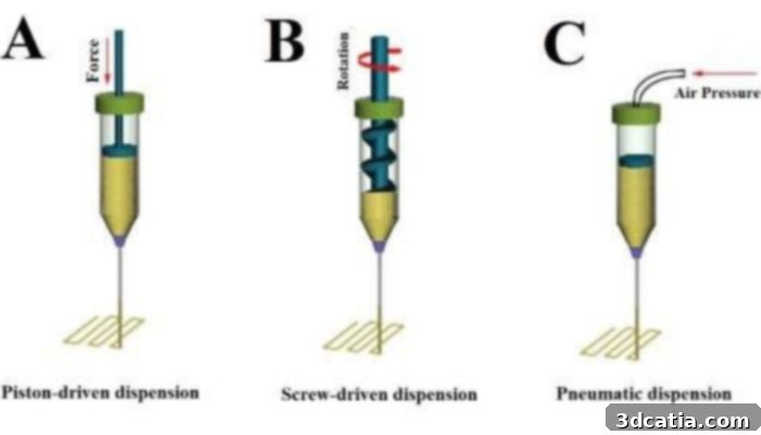
The team used a bioprinting method called pneumatic extrusion or dispension – C. Image credit: Advanced Nanobiomed Research on ResearchGate
Cultivating Growth: Microcalli Formation and Cell Regeneration
Beyond mere survival, a critical indicator of the success of any cell reproduction method is the ability of cells to resume normal biological functions, including division and differentiation. The NC State researchers meticulously assessed the production of microcalli from their viable bioprinted cells. Microcalli are small, unorganized masses of parenchyma cells—a common type of plant cell—which represent an initial stage of plant tissue regeneration and are crucial for processes like plant propagation and genetic transformation. The formation of these microcalli is a strong testament to the regenerative potential of the bioprinted cells.
The study observed that the cell cycle reentry within the bioprinted cells coincided with the activation of core cell cycle genes and genes intimately associated with cell regeneration pathways. This indicates that the bioprinting process not only kept the cells alive but also stimulated them to reactivate their fundamental growth mechanisms, effectively causing the bioprinted cells to restart their cell cycles and reproduce. This ability to manipulate and observe cellular regeneration in a controlled 3D environment is a significant advantage over traditional 2D cell cultures, which often lack the physiological relevance of a three-dimensional context.
A remarkable finding was that after just 14 days, an average of 90% of the bioprinted constructs had successfully formed 5-6 microcalli each. This high success rate unequivocally demonstrates that 3D bioprinted cells possess the inherent capability to undergo vigorous cell division and exhibit behavior consistent with cells produced through conventional, often more cumbersome, laboratory methods. This opens up unprecedented opportunities for studying plant development, stress responses, and genetic engineering in a precisely controlled and scalable manner, laying the groundwork for future advancements in plant biotechnology and sustainable agriculture.
Unveiling Cellular Identity and Environmental Responses in 3D Bioprinted Cells
Understanding cellular identity and its plasticity is fundamental to comprehending plant development and their adaptive capabilities. The NC State scientists took their investigation a step further by examining the cellular identity of the bioprinted root plant cells and the microcalli they formed. Using specific molecular markers to track protein expression, they observed dynamic changes in cell identity over time within the printed constructs. This ability to monitor cellular fate in a 3D environment provides invaluable insights that are difficult to obtain through conventional methods.
One of the most intriguing discoveries was the observation that the cells within the bioprinted structures induced genes responsible for the initiation or maintenance of stem cell identity within a mere three days of observation. Stem cells in plants, known as meristematic cells, are undifferentiated and possess the remarkable ability to develop into any type of specialized plant cell. This rapid induction of stem cell characteristics in bioprinted cells suggests a high degree of cellular plasticity and regenerative capacity, which is critical for tissue repair and growth in natural plant systems. This finding is particularly exciting because it implies that 3D bioprinting could be used to generate specific plant cell types or even complex tissues for various research and industrial applications.
To further evaluate the physiological robustness of their bioprinted cells, researchers also tested their responses to environmental stress. By applying high salinity conditions—a common abiotic stressor in agriculture—they assessed how the cells adapted. Unsurprisingly, both meristematic (non-specialized, rapidly dividing) and differentiated cells exhibited reduced viability under these stressful conditions. This finding is crucial because it validates that bioprinted plant cells respond to environmental cues in a manner similar to native plant cells, thereby enhancing the relevance of 3D bioprinting as a reliable platform for studying plant-environment interactions. This capability opens doors for future research into plant resilience, stress tolerance, and the development of crops better suited to challenging growing conditions.

Biological research could be improved with the use of 3D Bioprinting.
Broader Implications and Future Horizons for 3D Bioprinting in Plant Science
The compelling results from the North Carolina State University study carry profound implications for the trajectory of biological studies focused on plant cells. The demonstrated ability to produce plant cells with reasonable viability through 3D bioprinting positions this technology as a compelling alternative, and indeed an improvement, over traditional manual pipetting methods. As noted by corresponding co-author Professor Ross Sozzani, this innovative approach “provides better opportunity for high throughput processing and control over the architecture of the cells after bioprinting.” This control over cellular architecture is particularly transformative, allowing researchers to create complex, organized 3D structures that more closely mimic natural plant tissues than conventional 2D cultures, thereby providing a more biologically relevant experimental platform.
Enhanced Control and Scalability in Plant Research
The success in producing microcalli further underscores the vitality and regenerative capacity of these bioprinted cells, confirming their potential to re-enter the cell cycle and divide much like cells in living plants. This capacity is critical for detailed studies on plant development, growth regulators, and responses to various internal and external signals. Furthermore, the insights gained into cellular identity, particularly the rapid induction of stem cell characteristics and the observation of specific gene expression changes, are invaluable. As Lisa Van den Broeck, another key researcher, eloquently stated, “Bioprinted cells can take on the identity of stem cells; they divide and grow and express specific genes. When you bioprint, you print a whole population of cell types. We were able to examine the genes expressed by individual cells after 3D bioprinting to understand any changes in cell identity.” This ability to track and analyze individual cell responses within a heterogeneous bioprinted population is a powerful tool for dissecting complex cellular processes.
Looking ahead, the researchers suggest that future investigations could harness 3D bioprinting to explore cell viability, division, and identity within a ‘tunable environment.’ This concept of a tunable environment is revolutionary, implying the ability to precisely control and manipulate various physical and chemical parameters—such as nutrient composition, mechanical stiffness of the scaffold, pH, temperature, and even light exposure—in real-time. Such control would enable scientists to mimic specific environmental niches or stress conditions with unparalleled accuracy, facilitating deeper understanding of plant developmental pathways, disease resistance mechanisms, and how plants adapt to climate change.
Beyond Plant Cells: The Broad Spectrum of 3D Bioprinting
While this study focuses on plant cells, it’s important to remember that 3D bioprinting has already demonstrated its transformative potential across various biological projects. Notable examples include the ambitious development of medicinal organ growth for transplantation and the innovative production of meat alternatives to address sustainability and ethical concerns in food production. These diverse applications underscore the versatility and far-reaching impact of 3D bioprinting technology.
The application of 3D bioprinting to plant science is not merely a technical advancement; it represents a fundamental shift in how biological research can be conducted. Unlike some commercial applications of 3D printing, the value of 3D bioprinting in this context lies firmly in its contribution to pure scientific discovery. It offers a sophisticated, controlled, and high-throughput platform that can accelerate our understanding of plant biology, ultimately leading to breakthroughs in agriculture, pharmaceuticals, and environmental conservation. This is truly a very interesting and critical field for pushing the boundaries of scientific knowledge.
For those interested in delving deeper into the specifics of this pioneering research, the full report is readily available on Science.org (log-in required). Additionally, a concise summary can be found on the University’s official news blog HERE, offering an accessible overview of the key findings and their implications.
What are your thoughts on this groundbreaking study? Do you believe 3D bioprinting will fundamentally reshape the landscape of biological research, particularly in plant science? We encourage you to share your insights and predictions in the comments section below or engage with us on our social media platforms: LinkedIn, Facebook, and Twitter! Don’t miss out on the latest advancements in 3D printing; sign up for our free weekly Newsletter here to get news delivered directly to your inbox. You can also explore all our informative videos on our dedicated YouTube channel.
*Cover photo credit: Lisa Van den Broeck, NC State University