
Revolutionizing Oral Cancer Treatment: Personalized Drug Screening with 3D Printed Microfluidic Devices
The landscape of cancer treatment is continually evolving, with a strong focus on personalized medicine to deliver more effective therapies with fewer side effects. Imagine a world where scientists could predict how an individual cancer patient would respond to specific drugs *before* they even begin treatment. This isn’t a distant dream; it’s a rapidly advancing reality, largely due to innovative techniques like functional drug testing (FDT). This cutting-edge approach involves isolating tumor cells directly from a patient and culturing them in sophisticated environments to observe their reactions to various drug combinations. A pivotal tool in this research is the microfluidic device, engineered to meticulously mimic the intricate flow of fluids and cellular interactions within the human body. Leading this charge, the Indian Institute of Technology Hyderabad (IITH) has made remarkable strides by designing novel, 3D printed microfluidic devices specifically tailored for drug screening in oral cancer, aiming to develop a robust platform that provides unprecedented insights into drug-cancer cell interactions.
The conventional wisdom of “one-size-fits-all” cancer treatment is increasingly being challenged. Each patient’s cancer is unique, influenced by genetic makeup, tumor biology, and individual physiological responses. This inherent variability often leads to unpredictable outcomes, with some patients responding well to standard therapies while others experience minimal benefit or significant adverse reactions. Functional drug testing offers a powerful solution by shifting the paradigm from generalized treatments to highly personalized strategies. By directly testing patient-derived cancer cells against a spectrum of potential drugs, researchers can identify the most efficacious compounds for that specific individual, thereby maximizing therapeutic impact and minimizing unnecessary exposure to ineffective or toxic agents. This personalized approach holds the promise of transforming cancer care, making treatments more targeted, efficient, and ultimately, more successful.
The Precision of Microfluidics: Mimicking the Body’s Complex Environment
At the heart of IITH’s groundbreaking research lies the sophisticated application of microfluidic technology. Microfluidic devices are miniature laboratories on a chip, designed to handle and manipulate minute volumes of fluids (picoliters to microliters). Their ability to precisely control the flow and transport of substances at a micro-scale makes them ideal for simulating the physiological conditions experienced by cells within the body, such as nutrient delivery, waste removal, and drug exposure gradients. Unlike traditional 2D cell cultures in petri dishes, which offer a limited representation of the complex in vivo environment, microfluidic systems can replicate the dynamic and three-dimensional nature of biological systems more accurately. This includes maintaining specific temperatures, pH levels, and shear forces, all of which are critical for culturing cells in a manner that closely resembles their natural state within a living organism.
The innovative aspect of IITH’s work is their embrace of 3D printing for fabricating these intricate microfluidic devices. The IITH team meticulously designed their device for the study, selecting Formlabs’ clear resin as the optimal 3D printing material. This choice was not arbitrary; the resin was identified as providing the best environment for cultivating the specific oral cancer cells crucial to their research. Additive manufacturing, or 3D printing, offers unparalleled advantages in creating such complex structures. It allows for rapid prototyping, enabling researchers to iterate on designs quickly and efficiently. More importantly, it provides the precision and geometric freedom necessary to construct the intricate channels, chambers, and micro-structures required for effective microfluidic operation. This level of customization and detail would be incredibly difficult, if not impossible, to achieve with traditional manufacturing techniques, underscoring the transformative potential of 3D printing in biomedical research and personalized medicine.
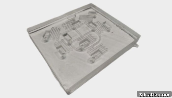
This image displays the custom-designed 3D printed microfluidic device created by IITH, highlighting the sophisticated engineering involved in replicating physiological conditions for oral cancer drug screening. (photo credit: Mehta, V., Vilikkathala Sudhakaran, S. et al., Journal of Nanobiotechnology, 2024)
Spheroids-on-a-Chip: A New Dimension in Tumor Modeling
To ensure the most accurate representation of patient tumors, IITH’s study involved three patients from whom biopsy samples were carefully collected. From these samples, oral tumor stem-like cells were isolated and further cultivated. Instead of growing these cells as a flat monolayer (2D culture), which fails to capture the true complexity of a tumor, the researchers encouraged them to form spheroids within the microfluidic device. Spheroids are three-dimensional, self-aggregating cellular structures that mimic the spherical clusters of cancer cells found in actual tumors. These structures are profoundly beneficial for research because they recreate the diverse cell populations, nutrient gradients, and oxygen levels that characterize in vivo tumors, thereby providing a far more physiologically relevant model than traditional 2D cultures. This innovative integration of spheroids within a microfluidic device led to the creation of what researchers term “spheroids-on-a-chip,” a powerful platform for advanced drug testing.
The IITH-designed 3D printed microfluidic device, functioning as this advanced “spheroids-on-a-chip” system, boasts an intricate two-layer network. This sophisticated architecture comprises serpentine loops specifically engineered for efficient and thorough mixing of various drug combinations, ensuring uniform exposure for the cultured cells. Complementing these mixing channels are cylindrical microwells, precisely sized and shaped for the optimal cultivation of the patient-derived spheroids. This thoughtful design allows researchers to simultaneously test multiple permutations of drugs. In this particular study, the team evaluated seven distinct combinations of three well-established oral cancer drugs: paclitaxel, 5-fluorouracil, and cisplatin. The ability to test combinations is critically important because tumors frequently develop resistance to single-agent therapies. By exploring synergistic drug combinations, scientists can identify strategies that overcome resistance mechanisms and offer a more potent therapeutic punch.
Personalized Insights and Overcoming Drug Resistance
The results of the IITH study underscored the immense value of personalized drug screening. For instance, the spheroids derived from patient 1 exhibited significant resistance to all tested drug combinations, indicating that standard treatments might prove ineffective for this individual. In stark contrast, spheroids from other patients responded positively to specific drug combinations or even mono-drug treatments. This variability highlights a crucial aspect of cancer biology: each tumor has a unique response profile. The study’s conclusion emphasized “the influence of tumor differentiation status on treatment responses, which has been rarely carried out in the previous reports.” This is a profound finding, suggesting that the developmental stage or degree of differentiation of cancer cells within a tumor significantly dictates its susceptibility to different drugs. By understanding this status, researchers can determine which drug combinations are most likely to yield beneficial outcomes for a given patient.
Further validating the robustness of their platform, the research team noted that “these characteristics also correlated with each patient’s diagnosis from clinical histopathological reports.” This direct correlation between the in vitro results from the 3D printed microfluidic device and actual clinical diagnoses provides powerful evidence of the platform’s predictive capability. It suggests that the spheroids-on-a-chip model accurately mirrors the biological behavior of patient tumors in their natural environment, thereby making it a reliable tool for forecasting treatment efficacy. This validation is critical for translating such research findings from the laboratory bench to the patient’s bedside, bringing us closer to truly personalized and effective cancer therapies.

This graphic illustrates the comprehensive study design and key findings from the IITH research, demonstrating the effectiveness of their 3D printed microfluidic platform in personalized drug screening for oral cancer. (Photo credit: Credit: Mehta, V., Vilikkathala Sudhakaran, S. et al., Journal of Nanobiotechnology, 2024)
Limitations and Future Directions in Precision Oncology
While the IITH study represents a significant leap forward in personalized drug screening for oral cancer, the researchers acknowledge certain limitations that pave the way for future advancements. The initial study primarily focused on the direct interaction between cancer spheroids and drugs. However, the tumor microenvironment is a complex ecosystem, comprising various other cell types such as fibroblasts, immune cells, and endothelial cells, all of which can profoundly influence drug responses and tumor progression. Future research aims to incorporate these additional cellular components into more complex models, thereby providing an even more comprehensive and physiologically relevant platform for drug testing. Furthermore, the study did not delve into the detailed mechanisms of drug absorption by the spheroids upon exposure – an area crucial for understanding drug pharmacokinetics at the cellular level. Addressing this limitation will involve developing advanced imaging techniques and analytical methods to track drug uptake and distribution within the 3D tumor models.
Scientists at IITH and other institutions are actively planning to address these limitations by developing increasingly sophisticated models. This includes integrating vascular structures within the microfluidic devices to simulate blood flow and nutrient exchange more accurately, as well as incorporating elements of the immune system to study immunotherapeutic responses. The goal is to create truly organ-on-a-chip or tumor-on-a-chip systems that can fully recapitulate the complexity of human biology. Such advancements will not only refine functional drug testing platforms but also contribute significantly to our understanding of cancer biology, drug resistance mechanisms, and the development of next-generation cancer therapies. This ongoing research underscores the dynamic nature of biomedical innovation and the relentless pursuit of more effective, patient-centric cancer treatments. To delve deeper into the intricate details of IITH’s pioneering study, interested readers are encouraged to consult the original, comprehensive report available here.
The Future is 3D Printed: Additive Manufacturing in Healthcare Innovation
The successful application of 3D printing in creating such advanced microfluidic devices highlights the transformative power of additive manufacturing in the biomedical field. Beyond just drug screening, this technology is revolutionizing areas from prosthetics and implants to tissue engineering and bioprinting. The ability to custom-design and rapidly produce intricate, biologically relevant structures opens up unprecedented avenues for research and clinical applications. As 3D printing technology continues to evolve, becoming even more precise, versatile, and accessible, its impact on personalized medicine and healthcare innovation will only grow. It promises a future where treatments are not only tailored to individual patients but also developed and tested on systems that closely mirror their own biology, leading to more predictable outcomes and significantly improved patient care.
What are your thoughts on the incredible potential of using additive manufacturing to create sophisticated microfluidic models for personalized cancer treatment? We invite you to share your perspectives and engage in the conversation! Let us know your insights in a comment below or connect with us on our LinkedIn, Facebook, and Twitter pages! Don’t forget to sign up for our free weekly newsletter here for the very latest 3D printing news delivered straight to your inbox! You can also find all our compelling videos on our YouTube channel, offering visual insights into the world of additive manufacturing.