
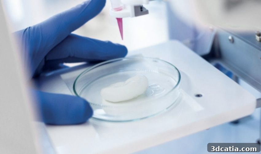
Revolutionizing Cancer Research: 3D Bioprinting Optimized by Cellular Automata Modeling
The rapid advancement of 3D bioprinting technology is fundamentally reshaping the landscape of biomedical research, opening up unprecedented avenues for understanding complex biological systems and developing innovative therapeutic strategies. A groundbreaking multi-university collaboration involving researchers from the University of Waterloo in Canada and the University of Swansea and the University of St Andrews in the UK has achieved a significant milestone in this exciting field. Their pioneering work centers on optimizing 3D bioprinting techniques for cancer cell studies by integrating traditional in-vitro experiments with sophisticated computational modeling. Specifically, the team conducted a detailed 11-day experimental study on 3D bioprinted cancer cells and then compared these empirical results with predictions from a cellular automata model. The success of their model in accurately simulating the behavior of the bioprinted structures offers a transformative approach, promising substantial savings in both time and financial resources for future experimental designs in cancer research and regenerative medicine.
Traditional two-dimensional (2D) cell cultures, while historically vital, often fail to replicate the complex and dynamic microenvironment found within living tissues and tumors. This limitation is particularly pronounced in cancer research, where the intricate interplay between cancer cells, the extracellular matrix, and various signaling molecules is crucial for understanding disease progression, metastasis, and therapeutic responses. Three-dimensional (3D) bioprinting emerges as a powerful solution to this challenge, enabling the creation of intricate scaffolds and cellular constructs that closely mimic the physiological architecture of native tissues. These advanced 3D models provide a more accurate and predictive platform for studying cancer biology, assessing drug efficacy, and investigating resistance mechanisms, offering insights that are often unattainable with conventional 2D methodologies. However, the process of developing, refining, and optimizing these 3D bioprinted models can be incredibly resource-intensive, highlighting the critical need for efficient validation and prediction tools like computational modeling.
Pioneering In-Vitro Experiments with Aggressive Breast Cancer Cells
In their meticulously designed in-vitro experimental method, the research team focused on MDA-MB-231 cells, an aggressive and extensively studied human breast cancer cell line. This particular cell line is favored in cancer research due to its highly metastatic characteristics and robust proliferative capabilities, making it an excellent model for investigating the dynamics of invasive breast cancer and potential therapeutic interventions. These specific cancer cells were precisely bioprinted within a specialized hydrogel, which served as a crucial biological scaffold. This hydrogel was engineered using a sophisticated bioink composed of a mixture of gelatin and alginate. The choice of these two biopolymers was deliberate and strategic. Both gelatin, a denatured form of collagen, and alginate, a natural polysaccharide derived from brown algae, are highly valued for their exceptional biocompatibility and their remarkable structural similarities to the native extracellular matrix (ECM). The ECM is a vital non-cellular component that provides structural support to cells and tissues, mediates cell-cell communication, and influences various cellular behaviors, including growth, migration, and differentiation. By closely mimicking the native ECM, the researchers ensured that the bioprinted cancer cells were cultured in an environment that faithfully replicated their natural biological surroundings, thereby promoting more accurate and physiologically relevant experimental outcomes.
The viability and proliferation of the bioprinted cellular structures were rigorously monitored over an extended period of 11 days. This specific observation window was essential for assessing the long-term health, functionality, and growth dynamics of the cancer cells within the complex 3D construct. The results of this careful monitoring were highly encouraging, revealing an impressive cell viability rate of 96 ± 2% after the 11-day period. Such a high survival rate is a clear indicator of the success and robustness of the bioprinting technique employed, as well as the suitability of the chosen bioink and the overall scaffold design. It unequivocally demonstrated that the bioprinted structure possessed sufficient porosity, which is paramount for the effective permeation and diffusion of essential nutrients, such as glucose, and crucially, oxygen. Adequate oxygen and nutrient supply are critical for sustaining cellular respiration and overall metabolic activity, particularly in dense 3D tissue models where cells deeper within the construct can experience hypoxia or nutrient deprivation. The sustained viability of the vast majority of the cells confirmed that the engineered microenvironment was highly conducive to supporting prolonged cell growth and function, thereby establishing a robust and reliable platform for subsequent, in-depth investigations into cancer cell behavior and their responses to various therapeutic agents.
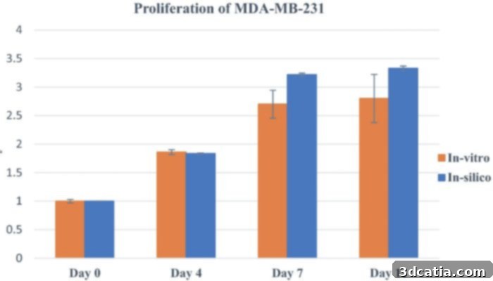
This graph vividly illustrates the comparative proliferation rates of experimental cells within the mathematical model against the actual results obtained from physical experiments. The Y-axis quantifies the number of proliferating cells, clearly demonstrating the remarkable accuracy and predictive power of the computational model. (Photo credit: Scientific Reports (Sci Rep))
Leveraging In-Silico Modeling: The Power of Cellular Automata
Complementing the empirical data obtained from in-vitro experiments, the researchers strategically employed in-silico modeling to predict and understand the complex dynamics of the bioprinted cancer cells. The term “in-silico,” a pseudo-Latin phrase, cleverly mirrors “in-vitro” and “in-vivo,” referring specifically to experiments or research conducted entirely by computer simulation or through computational models, drawing its name from the silicon chips integral to modern computing. For this pivotal research, the scientists utilized cellular automata modeling – a powerful computational technique that simulates complex systems by defining simple rules for cells arranged on a regular grid. In this paradigm, each “cell” within the grid, representing a biological cell or a cluster of cells, interacts dynamically with its neighboring cells and changes its state based on a predetermined set of rules. This enables the simulation of various biological processes, including cell proliferation, migration, differentiation, and even programmed cell death. A crucial aspect of their approach was the derivation of most parameters for the cellular automata model directly from the extensive experimental data gathered during the in-vitro studies. This data-driven parameterization was essential for grounding the model in empirical reality, significantly enhancing its predictive accuracy and biological relevance.
To further enhance the reliability and predictive capabilities of their sophisticated computational model, a comprehensive in-vitro assay was meticulously performed. This detailed examination of the bioprinted cells allowed researchers to precisely determine their composition, individual characteristics, and nuanced responses under a variety of controlled conditions. The invaluable insights garnered from these rigorous assays were then seamlessly integrated back into the cellular automata model, facilitating an iterative process of refinement and precise calibration. This meticulous validation protocol was absolutely critical, ensuring that the model was not merely theoretically sound but also possessed the robust capability to accurately reflect the complex biological realities observed in the laboratory setting. The final results of this advanced integration of experimental and computational methodologies were exceptionally promising: the 3D bioprinting optimization achieved through this innovative modeling technique can indeed be reliably utilized to design future experiments with an unprecedented degree of confidence. This revolutionary capability implies that researchers can now potentially achieve desired experimental outcomes and gather robust, meaningful data without the necessity of conducting numerous, often time-consuming, and resource-intensive preliminary in-vitro assays. Most critically, the simulated data consistently and accurately replicated the experimental results for both cell viability and proliferation, demonstrating a remarkable concordance. This unprecedented level of correlation between the computational model and physical experiments powerfully underscores the model’s reliability and its profound potential to revolutionize experimental design and accelerate discovery in the critical fields of bioprinting and cancer research.
Transforming Research: Accelerating Discovery and Enhancing Efficiency
The successful validation of this integrated computational and experimental approach carries profound implications for the entire biomedical research ecosystem. By conclusively demonstrating that a cellular automata model can accurately predict the complex behavior of 3D bioprinted cancer cells, this research provides a powerful and indispensable tool for unprecedented experimental optimization. Instead of embarking on countless costly and time-consuming physical experiments to determine optimal bioprinting parameters, bioink compositions, or cellular responses to various stimuli, researchers can now confidently leverage this validated computational model. This allows for rapid iteration, testing of multiple hypotheses, and exploration of various experimental conditions in a virtual environment, significantly reducing the dependency on extensive and often laborious laboratory work. The primary and most immediate benefits include substantial time savings, as complex simulations can be executed and analyzed far more rapidly than biological experiments, and considerable monetary savings, by minimizing the consumption of expensive reagents, specialized cell lines, and sophisticated laboratory equipment. Furthermore, this innovative approach aligns perfectly with modern ethical considerations in research by potentially reducing the number of necessary in-vitro assays and, consequently, diminishing the reliance on animal testing in early preclinical research stages. This transformative shift towards “virtual experimentation” liberates invaluable resources, enabling scientists to dedicate their efforts to more complex, impactful scientific questions and to dramatically accelerate the pace of discovery in critical areas such such as cancer treatment, drug development, and regenerative medicine.
The Broader Impact of 3D Bioprinting: From Eye Tissues to Space Exploration
Beyond its immediate application in cancer research optimization, 3D bioprinting continues to emerge as a field of immense transformative potential, holding critical applications across regenerative medicine, advanced tissue engineering, and pharmaceutical development. Its remarkable capability to construct intricate biological architectures layer by layer is unlocking previously unimaginable possibilities for therapeutic interventions and disease modeling. A prime example of this groundbreaking utility is its application in creating complex eye tissues. This advancement is profoundly significant for addressing debilitating conditions such as age-related macular degeneration, corneal damage, glaucoma, and various retinal diseases. It offers a beacon of hope for developing innovative therapeutic strategies, potentially leading to sight restoration, and provides invaluable platforms for creating accurate disease models for high-throughput drug screening specifically tailored to ocular conditions. Expanding its reach even further, 3D bioprinting is transcending terrestrial boundaries and venturing into extraterrestrial environments, as strikingly evidenced by the Ugandan government’s recent groundbreaking initiative to launch a 3D bioprinter into space. The paramount objective of such pioneering missions is to conduct crucial tissue experiments under conditions of zero gravity, meticulously exploring how microgravity environments uniquely affect cellular growth, differentiation, and complex tissue formation. Such insights could have profound implications for long-duration human space travel, the understanding of fundamental biological processes unfettered by gravitational influence, and even the potential for manufacturing biopharmaceuticals or tissues in space.
The present experiment, which ingeniously focuses on optimizing 3D bioprinting through the development and rigorous validation of a robust computer model against empirical experimental results, is exceptionally significant for the future of biomedical science. It represents a pivotal leap towards achieving truly predictive biology, enabling the accurate forecasting of complex cellular activities and their responses within dynamically bioprinted constructs. This unprecedented predictive capability is instrumental in minimizing the need for extensive, often repetitive, experimental evaluations, thereby maximizing research efficiency and substantially reducing resource expenditure. The implications of this research extend broadly to accelerating the development of novel cancer therapies, engineering functional and transplantable tissues for regenerative purposes, and creating more accurate, physiologically relevant disease models that can revolutionize drug discovery and personalized medicine. By providing a reliable, validated methodology to simulate and predict biological outcomes, this innovative research empowers scientists to design more effective, targeted, and data-rich experiments, thereby driving innovation forward while simultaneously realizing monumental time and cost savings. This exemplary multi-university collaboration powerfully underscores the profound potential of interdisciplinary research in effectively tackling humanity’s most complex scientific challenges and rapidly advancing the frontiers of biomedical engineering and clinical translation.

Bioprinting demonstrates remarkable versatility, with applications spanning both the medical and botanical fields. An illustrative example is this innovative 3D-printed blend of algae and cellulose, a creation from the Delft University of Technology (TU Delft), showcasing the immense potential for sustainable biomaterials and novel biological structures. (Photo credit: TU Delft)
This pioneering research, extensively detailed in a comprehensive scientific paper accessible HERE, signifies a critical advancement in the sophisticated integration of computational modeling with cutting-edge additive manufacturing techniques like 3D bioprinting. The ability to accurately predict complex biological outcomes in a virtual environment dramatically reduces the need for extensive, often trial-and-error based, experimentation in the laboratory. This not only makes the entire research process significantly faster and more economical but also enhances its ethical foundation by minimizing the use of biological resources. Such synergy between advanced simulation and experimental validation is absolutely vital for addressing the multifaceted and complex challenges inherent in modern medicine, particularly in the critical realms of personalized cancer treatment, the development of functional tissue grafts for transplantation, and the creation of advanced organ-on-a-chip systems for drug discovery. As 3D bioprinting technologies continue their rapid evolution, synergistically combined with increasingly sophisticated in-silico models, we can anticipate a future where the routine creation of functional human organs and the implementation of highly personalized therapeutic strategies become an achievable reality, fundamentally transforming healthcare and human well-being as we know it.
What are your thoughts on this groundbreaking 3D bioprinting experiment and the profound potential of combining advanced computational modeling with experimental biology? We sincerely invite you to share your valuable insights and perspectives in a comment below or to join the vibrant conversation on our Linkedin, Facebook, and Twitter pages! Don’t miss out on staying abreast of the latest advancements, news, and innovations in the dynamic world of additive manufacturing; make sure to sign up for our free weekly Newsletter here, conveniently delivered straight to your inbox. You can also explore all our engaging videos, insightful interviews, and in-depth content on our dedicated YouTube channel, where we regularly feature the cutting edge of 3D printing technology and its applications.
*Cover photo credits: 3Dnatives