
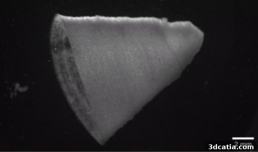
Harvard’s Bioprinting Breakthrough: Engineering a Functional 3D Printed Heart Ventricle with Fiber-Infused Ink
Cardiovascular diseases, collectively known as heart disease, stand as a formidable global health challenge, consistently ranking among the leading causes of mortality worldwide. The staggering statistics underscore the urgency of advanced medical research and innovative therapeutic approaches. According to the Centers for Disease Control and Prevention (CDC), the United States alone witnesses a death from cardiovascular disease approximately every 33 seconds. In 2021, these diseases were responsible for an alarming one in five deaths, affecting an estimated 695,000 individuals. This pervasive threat necessitates continuous breakthroughs in treatment and prevention, making every scientific advancement in cardiac health critically important. It is against this backdrop that recent developments from Harvard scientists offer a beacon of hope, leveraging the cutting-edge technology of bioprinting to create what could be a foundational step towards solving complex cardiovascular challenges.
This pivotal achievement involves the use of a specially formulated, fiber-infused ink, which has enabled researchers to successfully 3D print a functional heart ventricle. Remarkably, this bioprinted structure is capable of mimicking the intricate beating patterns characteristic of a human heart. Such a development marks a significant leap forward in the field of regenerative medicine, moving beyond theoretical models to tangible, functional biological constructs. The implications for understanding heart function, testing new drugs, and eventually creating transplantable organs are profound, potentially reshaping the landscape of cardiovascular care and addressing one of the most pressing health crises of our time.
Addressing the Critical Organ Shortage with Bioprinting Innovation
The concept of using bioprinting to construct living organs is not entirely new, but the journey towards creating viable, transplantable structures has been fraught with challenges. The current reality of organ donation highlights an acute and persistent shortage: while the Organ Procurement and Transplantation Network reported over 42,000 transplants in 2022, the demand far outstrips supply, leading to tragic consequences. An estimated 17 individuals succumb each day while patiently awaiting a life-saving organ transplant. This grim statistic underscores the desperate need for alternative solutions. Bioprinting, which involves precisely depositing living cells and biomaterials layer by layer to create functional tissues and organs, has emerged as a serious contender to bridge this critical gap.
However, despite extensive research and significant investments, establishing the long-term viability and complexity required for full-scale organ transplantation has proven difficult. Previous efforts often struggled to replicate the intricate architectures and dynamic functions of natural organs, particularly the rhythmic, coordinated contractions of a heart. The Harvard breakthrough, by enabling the creation of tissue that can genuinely beat and pump fluid, represents a monumental stride. It brings us considerably closer not only to developing accurate 3D printed tissues for advanced research and drug screening but also to the ambitious goal of bioprinting entire, fully functional organs for therapeutic use. This innovation provides a new pathway for scientists to overcome previous limitations, pushing the boundaries of what is possible in bioengineering.
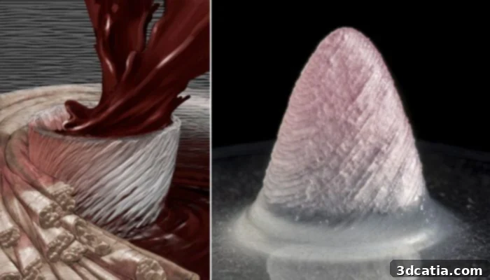
Suji Choi, a distinguished research associate at the Harvard John A. Paulson School of Engineering and Applied Sciences (SEAS) and the first author on the groundbreaking paper, eloquently explains the unique advantage of their developed material: “People have been trying to replicate organ structures and functions to test drug safety and efficacy as a way of predicting what might happen in the clinical setting. FIG ink is capable of flowing through the printing nozzle but, once the structure is printed, it maintains its 3D shape. Because of those properties, I found it’s possible to print a ventricle-like structure and other complex 3D shapes without using extra support materials or scaffolds.” This inherent structural integrity of the fiber-infused gel (FIG) ink after deposition is what sets this method apart, streamlining the bioprinting process and opening new avenues for creating intricate biological architectures with unprecedented ease and precision.
Engineering a Working 3D Printed Heart: The Science Behind the Success
The detailed methodology and exciting results of this research were recently published in the prestigious journal Nature Materials. The team of brilliant researchers from the Harvard John A. Paulson School of Engineering and Applied Sciences (SEAS) has pioneered the development of a novel hydrogel ink, ingeniously infused with gelatin fibers. This innovative composite material, aptly named fiber-infused gel (FIG) ink, is at the heart of their breakthrough. The unique properties of FIG ink have been harnessed to enable the precise 3D printing of a functional heart ventricle, a structure that not only mimics the anatomical form but also replicates the vital beating action of a real human heart.
The process begins with the FIG ink being extruded through a printing nozzle. What distinguishes this ink from many predecessors is its remarkable ability to immediately retain its complex 3D shape upon deposition. This critical characteristic is directly attributable to the inclusion of the gelatin fibers within the hydrogel matrix. As Suji Choi elucidated, this property eliminates the need for cumbersome and often problematic extra support materials or scaffolds that are typically required in traditional bioprinting to prevent printed structures from collapsing. The structural integrity provided by the fibers allows for the creation of intricate ventricle-like geometries with high fidelity. Furthermore, the Harvard team demonstrated a sophisticated level of control over the printing direction, a crucial factor that allowed them to precisely influence and align the heart muscle cells within the printed tissue. This directed alignment is essential for achieving coordinated contractions, mirroring the highly organized architecture of native heart tissue.
Once electrical stimulation was judiciously applied to the bioprinted ventricle, it initiated a synchronized wave of contractions. These contractions occurred in precise alignment with the pre-established direction of the embedded fibers and the aligned muscle cells, effectively replicating the pumping action of a natural heart. While the current iteration is still a simplified and miniaturized model compared to a full human organ, its functionality is groundbreaking. The researchers are actively engaged in developing more life-like heart tissues, aiming for structures with greater thickness and increased muscular complexity to more closely approximate in vivo conditions. Even in its current state, this 3D printed ventricle showcases significant performance enhancements, demonstrating the capacity to pump between 5 to 210 times more fluid volume than any previous 3D printed heart models. This substantial improvement in pumping efficiency represents a pivotal step, paving the way for the future development of more advanced cardiac components, including functional heart valves, complex dual-chambered miniature hearts, and even comprehensive models for studying cardiac diseases and drug responses.
The Broader Vision: FIG Ink and the Future of Regenerative Therapeutics
The significance of the FIG ink technology extends far beyond the creation of simple ventricle models. Kevin “Kit” Parker, the Tarr Family Professor of Bioengineering and Applied Physics and Head of the Disease Biophysics Group at SEAS, also a senior author on the paper, offers a broader perspective on this advancement. He emphasizes that FIG ink is just one tool in a growing arsenal: “FIGs are but one tool we have developed for additive manufacturing. We have other methods in development as we continue our quest to build human tissues for regenerative therapeutics. The goal is not to be tool driven – we are tool agnostic in our search for a better way to build biology.” This statement highlights a fundamental philosophy driving the research: a relentless pursuit of innovative methods to construct biological tissues, with an ultimate goal of translating these lab-based creations into practical, life-saving regenerative therapies.
The quest to “build biology” encompasses a wide range of applications, from creating organoids for disease modeling and drug discovery to developing transplantable tissues that can repair or replace damaged organs. The ability to precisely control the architecture and functionality of bioprinted tissues, as demonstrated with the FIG ink, is a critical component of this ambitious vision. It allows scientists to create more accurate in vitro models of human physiology, reducing reliance on animal testing and accelerating the development of new treatments for conditions like heart failure, congenital heart defects, and various forms of cardiovascular disease. The future potential includes personalized medicine, where patient-specific tissues could be engineered to perfectly match an individual’s unique biological makeup, minimizing rejection risks and optimizing therapeutic outcomes.
This Harvard breakthrough represents a significant milestone in the ongoing revolution of bioprinting and regenerative medicine. While challenges remain in scaling up these technologies to produce full-sized, long-lasting organs suitable for human transplantation, the functional heart ventricle printed with FIG ink provides tangible proof of concept and invaluable insights into the complexities of biofabrication. It moves us closer to a future where organ shortages might become a thing of the past and where complex diseases can be understood and treated with unprecedented precision. You can delve deeper into the specifics of this ground-breaking research, including the detailed method for fabricating the innovative fibers, by accessing the official press release HERE.
What are your thoughts on this latest research from Harvard? Do you believe that fiber-infused ink, like the groundbreaking FIG ink, will significantly expand the applications for bioprinting, particularly in the creation of functional 3D printed hearts and other complex organs? Share your insights and perspectives in a comment below or join the conversation on our LinkedIn, Facebook, and Twitter pages! Don’t miss out on the latest advancements and news in the dynamic world of 3D printing; remember to sign up for our free weekly Newsletter here, delivering the freshest updates straight to your inbox! Additionally, you can explore all our engaging videos and in-depth content on our dedicated YouTube channel.