
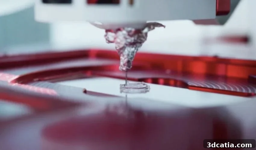
Revolutionizing Respiratory Research: McMaster University Develops Groundbreaking 3D Bioprinting Bioink for Lung Tissue
The field of 3D bioprinting continues its rapid evolution, emerging as an exceptionally promising tool in the advancement of personalized medicine and regenerative therapies. This innovative technology holds the potential to transform how we approach complex medical conditions, offering bespoke solutions tailored to individual patient needs. Recognizing this immense potential, a dedicated team of researchers at McMaster University in Ontario has achieved a significant breakthrough: the development of a novel bioink specifically engineered to accurately mimic the intricate mechanical and structural properties of human lung tissue. This isn’t just another step forward; it’s a leap. This new bioink allows for the printing of sophisticated tissues that are not only structurally sound but also functionally dynamic, capable of contracting and “breathing” much like natural lungs. Initially, the primary focus of this groundbreaking research is to accelerate medical discovery and facilitate the development of more effective treatments for prevalent respiratory diseases. However, looking towards the future, scientists envision direct clinical applications, including the repair of damaged lungs through transplantation or implantation in patients suffering from debilitating conditions such as Chronic Obstructive Pulmonary Disease (COPD) or pulmonary fibrosis, offering a beacon of hope for millions.
What sets this pioneering material apart from conventional options is its remarkable stability and versatility. Many existing bioinks often necessitate low temperatures for the printing process, which can sometimes compromise the viability of embedded cells and lead to structural degradation once the tissue is formed. Furthermore, these materials frequently struggle to maintain their intended shape and integrity post-printing, particularly when confronted with the dynamic environment of the human body. In stark contrast, McMaster’s innovative bioink is designed to retain complex, high-resolution structures with exceptional fidelity, all while remaining perfectly stable at physiological body temperature. This crucial characteristic ensures that the printed tissues can function optimally within a biological context, making them invaluable for both research and potential clinical use. The project, which received vital financial backing from McMaster University in 2024, has already catalyzed the creation of a promising startup, Tessella Biosciences. This new venture has quickly gained traction, securing its first customers and receiving overwhelmingly positive feedback, underscoring the immediate relevance and profound impact of this scientific advancement on the medical and biotechnological landscape.
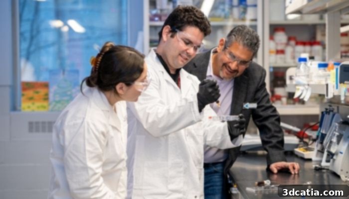
From left to right, David González Martinez and José Morán-Mirabal working with bioink for bioengineered lung tissue (Photo credit: Georgia Kirkos, McMaster University)
Addressing Critical Challenges: Why Develop a Bioink for Lung Tissue?
The impetus behind this ambitious project stemmed from a long-standing and significant constraint in respiratory disease research. Jeremy Hirota, Associate Professor of Medicine at McMaster University and a co-founder of the Tessella Biosciences startup, articulated the core challenge: the immense difficulty in accurately recreating realistic cellular environments necessary for a comprehensive study of complex conditions such as COPD and pulmonary fibrosis. These diseases, characterized by progressive and often irreversible damage to lung tissue, affect millions globally, yet our understanding of their precise mechanisms and our ability to develop effective therapies are often hindered by inadequate research models. Traditional research methods, while foundational, often fail to capture the dynamic, three-dimensional nature of living organs. Hirota emphasized this disparity, stating, “Lungs breathe. They open and close with every breath we take. But 95 to 99 per cent of the research we do in the lab is done on hard plastic dishes, whether it’s a petri dish or a tissue culture plate. It doesn’t take a scientist to understand that this hard plastic is not what your lungs are.” This highlights a critical disconnect: studying a highly elastic, constantly moving organ on a rigid, static surface inherently limits the insights gained into disease progression, drug efficacy, and cellular responses.
To bridge this crucial gap between static lab models and dynamic biological reality, Professor Hirota initiated a collaborative effort with José Moran-Mirabal, a distinguished professor in the Department of Chemistry and Chemical Biology, and David Gonzalez Martinez, a talented doctoral student. This interdisciplinary collaboration, bringing together expertise in medicine, materials science, and bioengineering, was instrumental in forging the path toward a truly revolutionary solution. The outcome of their combined efforts is a specially formulated bioink, meticulously designed to reproduce the quintessential elasticity and stretchability that define healthy lung tissue. This achievement is attributed to its precisely adapted composition and rheological properties, which allow the printed structures to mimic the mechanical movements of natural lungs. The researchers proudly describe this novel bioink as a “plug-and-play” solution, a testament to its remarkable compatibility with a wide array of currently available 3D bioprinters. This ease of integration is a significant advantage, eliminating the need for specialized equipment or complex adjustments, thereby making advanced tissue engineering more accessible to a broader scientific community. This innovative material enables the rapid production of complex, three-dimensional tissue structures, often in less than an hour, all while maintaining exceptionally high resolution and structural integrity.
The immediate implications and potential applications of this advanced bioink are vast and transformative. While it already demonstrates immense promise for enhanced lung modeling and significantly improved drug toxicity and response testing, the McMaster team is actively exploring an even wider spectrum of future clinical applications. In the short to mid-term, these include the potential for manufacturing highly realistic and functional skin grafts, which could revolutionize treatment for severe burn victims by providing more effective and customizable wound healing solutions. More directly related to its primary purpose, the bioink could also be used to produce functional fragments of lung tissue for targeted repair or transplantation procedures. This could offer a vital alternative for patients with localized lung damage, potentially reducing the need for full organ transplants and mitigating associated risks. Such developments could dramatically improve the quality of life for individuals suffering from chronic lung diseases. Beyond these immediate horizons, researchers harbor the ambitious long-term vision of biologically printing entire, fully functional organs. This aspiration, often referred to as the “holy grail” of 3D bioprinting, represents a monumental goal in the field. However, the team candidly acknowledges that achieving this ultimate objective still faces significant hurdles. These challenges are multifaceted, encompassing profound scientific complexities, such as ensuring vascularization and innervation of large printed organs, maintaining long-term cell viability, and mitigating immune rejection. Additionally, substantial regulatory hurdles must be overcome to ensure the safety, efficacy, and ethical deployment of such advanced bioprinted organs in clinical settings. Despite these formidable obstacles, the progress made by McMaster University’s team signals a profound shift in our capacity to understand, treat, and ultimately repair complex human tissues and organs, marking a pivotal moment in the journey towards true regenerative medicine and personalized healthcare.
What are your thoughts on this groundbreaking new bioink for lung tissue and its potential to revolutionize respiratory medicine? Let us know your insights and opinions in a comment below or join the conversation on our LinkedIn, Facebook, and Twitter pages! Don’t forget to sign up for our free weekly newsletter here to receive the very latest 3D printing news directly to your inbox! You can also find all our engaging videos on our dedicated YouTube channel. For those interested in more medical and dental 3D printing news, explore our specialized page HERE.
*Photo Credits: McMaster University