
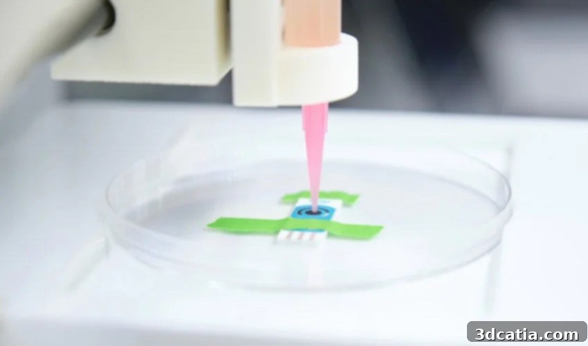
Revolutionizing Organ Transplantation: Microfluidic Bioprinting at Stevens Institute of Technology
The advent of 3D bioprinting has opened exciting new frontiers in regenerative medicine, promising solutions to some of humanity’s most pressing health challenges. At the cutting edge of this innovation is a pioneering team of researchers at the Stevens Institute of Technology in the United States. They are currently focused on developing an advanced bioprinting method rooted in microfluidics. This sophisticated technique involves the precise manipulation of fluids at the micrometer scale, offering an unprecedented level of control and detail. The significance of this project lies in its potential to enable researchers to work at a much finer resolution than previously possible, facilitating the accurate creation of any type of human tissue. By reproducing the intricate biological characteristics of human body cells with remarkable precision, this technology holds immense promise for advancing critical medical fields, most notably organ transplantation.
Addressing the Dire Organ Shortage Crisis with Innovative Solutions
The global shortage of organs for transplantation remains a severe public health crisis. In the United States alone, the numbers are stark: according to the Division of Transplantation (DoT) of the Health Resources and Services Administration at the U.S. Department of Health and Human Services, a staggering 105,940 individuals are currently on the national transplant waiting list. This critical scarcity of available organs leads to tragic outcomes for thousands across the country, highlighting an urgent need for revolutionary alternatives to traditional donation methods. This is where 3D bioprinting, a technology capable of reproducing customized cellular structures, steps in as a beacon of hope.
Bioprinting has the inherent capability to engineer complex biological constructs, from personalized skin grafts for burn victims to intricate organs. While the dream of routinely 3D printing a fully functional human heart or a kidney ready for transplant might still be a long-term goal, the rapid advancements being made, particularly by Professor Robert Chang’s team at Stevens Institute, clearly indicate that significant and tangible progress is being made. These developments are bringing us closer to a future where customized organs could be created on demand, potentially saving countless lives.
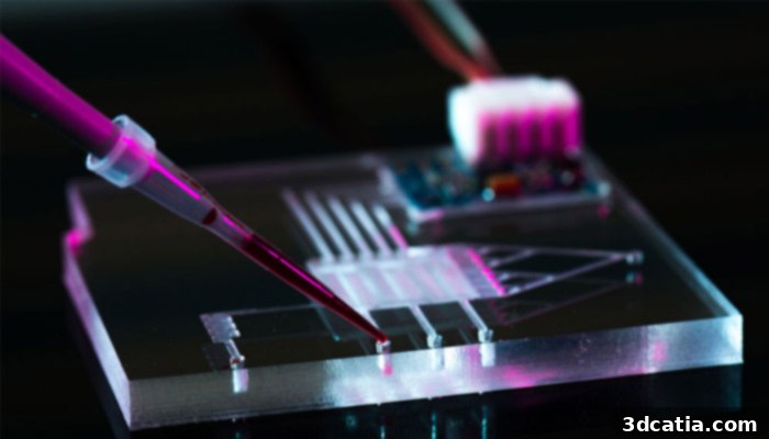
Microfluidics is a science that manipulates fluids with characteristics in the micrometer range, essential for achieving high-resolution bioprinting.
The Microfluidics Advantage: Superior Precision for Tissue Engineering
The pioneering research led by Professor Robert Chang at the Stevens Institute of Technology offers a significant leap forward in bioprinting technology. Its distinct advantage lies in its reliance on microfluidics, a method that provides far greater control and resolution compared to many existing bioprinting approaches. A majority of other 3D bioprinters currently available primarily utilize extrusion-based processes. These systems typically extrude bio-inks—hydrogels infused with living cells—layer by layer, with a common layer thickness of approximately 200 microns. While effective for certain applications, this resolution often proves insufficient for replicating the extremely fine and complex micro-architectures found within native human tissues.
Microfluidic bioprinting, however, drastically enhances this precision. By leveraging microfluidic principles, it becomes possible to achieve printing resolutions down to just a few tens of microns. This remarkable reduction in scale is fundamentally critical because it allows the bioprinted structures to much more closely mimic the actual dimensions and spatial arrangements of individual cells and their surrounding extracellular matrix within the human body. This unprecedented level of detail is crucial for creating functional and biologically accurate tissues that can effectively integrate and perform within a living system.
Overcoming the Resolution Barrier: Professor Chang’s Vision
Professor Robert Chang emphasizes the importance of this fine control: “Creating new organs to order and saving lives without the need for a human donor will be an immense benefit to healthcare. However, reaching that goal is tricky because printing organs using “bio-inks” — hydrogels laden with cultured cells — requires a degree of fine control over the geometry and size of printed microfibers that current 3D printers simply can’t achieve.” His insights underscore the limitations that traditional bioprinting methods face when attempting to replicate the complex architecture necessary for viable organs. The Stevens team’s microfluidic approach directly tackles this challenge, enabling the construction of intricate microfibers and cellular arrangements that closely mirror natural biological structures, thereby overcoming a significant hurdle in the quest for functional organ bioprinting.
The Engine of Innovation: Stevens Institute’s Computational Model
To harness the full potential of microfluidic bioprinting, Professor Chang’s team has engineered a sophisticated computational model designed for a microfluidic print head. This model acts as the intelligent core of their system, providing unparalleled control over crucial printing parameters such as flow speed, fluid dynamics, and pressure. This digital precision allows researchers to meticulously modify the geometries and material properties of the bioprinted structures in real-time, enabling a level of customization and accuracy previously unattainable. By understanding and predicting fluid behavior at the micro-scale, the model ensures that bio-inks are deposited exactly where and how they are needed.
A truly groundbreaking capability facilitated by this computational model is the ability to seamlessly mix multiple bio-inks simultaneously. This means that various types of cells, each with their unique biological functions and growth requirements, can be strategically combined and precisely patterned within a single bioprinted construct. This multi-material approach is indispensable for creating complex organs, which are inherently composed of diverse cell populations arranged in highly specific architectures. For example, to print a functional liver, a variety of cell types—hepatocytes, endothelial cells, stellate cells—must be integrated and organized correctly. The ability to choreograph the deposition of these diverse cellular components with such precision represents a monumental stride forward in achieving biologically accurate and functional tissue engineering.
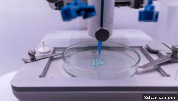
Current 3D bioprinters are mainly based on an extrusion process, which offers less precise resolution compared to advanced microfluidic techniques (photo credits: Département06-Xavier Giraud).
Building the Future: From Tissues to Complex Organs
The early achievements of Professor Chang’s team are already demonstrating the significant potential of their microfluidic bioprinting method. They have successfully printed bladders using 3D printed scaffolds, showcasing the viability of their approach for creating complex organ shapes. However, their ambition extends far beyond simple structures. By precisely combining and arranging multiple bio-inks, they aim to go much further, engineering intricately functional tissues and organs that closely mimic the biological complexity of native human counterparts. This involves not just structural accuracy but also the ability to integrate crucial biological functions, such as vascularization for nutrient supply and waste removal, which are essential for long-term viability.
Robert Chang articulates this expansive vision: “Being able to operate at this scale, while precisely mixing bio-inks, makes it possible for us to reproduce any tissue type. This technology is still so new that we don’t know precisely what it will enable. But we know it will open the door to creating new structures and important new types of biology.” This powerful statement highlights the immense, yet still largely uncharted, territory that microfluidic bioprinting is opening. It suggests a future where not only existing tissues and organs can be replicated, but entirely new biological structures or therapies, previously unimaginable, could be engineered.
Transformative Impact on Healthcare and Beyond
The implications of this cutting-edge research extend far beyond merely addressing the organ shortage. Successfully developing techniques to create customized, fully functional human tissues and organs would revolutionize numerous facets of healthcare and biomedical research. Imagine the possibilities: personalized organs perfectly matched to a patient’s immune system, virtually eliminating the debilitating risk of organ rejection that currently plagues transplant recipients. This advancement alone could dramatically improve patient outcomes and quality of life.
Furthermore, microfluidic bioprinting opens new avenues for creating accurate human disease models in vitro. These models would allow researchers to study disease progression and test new drug therapies with unprecedented fidelity, potentially reducing reliance on animal testing and accelerating the discovery of life-saving medications. The precision offered by this technology could also lead to groundbreaking advancements in tissue repair and regeneration. Damaged tissues could be precisely reconstructed with biologically accurate scaffolds and cellular compositions, offering hope for treating conditions currently considered untreatable, such as severe neurological damage or complex musculoskeletal injuries. The pioneering work at Stevens Institute of Technology is not just about advancing 3D printing; it’s about fundamentally reshaping the landscape of medical treatment, drug development, and our understanding of human biology itself.
For those eager to learn more about the remarkable progress made by Professor Chang’s team and other innovations in this field, we invite you to visit the Stevens Institute website HERE. We encourage you to share your insights and opinions on this transformative microfluidic bioprinting method. Feel free to leave a comment below or join the vibrant discussion on our Linkedin, Facebook, and Twitter pages! To stay updated with the latest advancements in additive manufacturing, don’t forget to sign up for our free weekly Newsletter here, delivering the most relevant 3D printing news directly to your inbox. Additionally, explore all our engaging video content on our YouTube channel, showcasing more incredible breakthroughs shaping the future of 3D printing.