
Revolutionizing Healthcare: The Transformative Power of 3D Bioprinting in Personalized Medicine and Beyond
3D bioprinting stands at the forefront of medical innovation, poised to revolutionize healthcare by offering groundbreaking solutions that range from advanced tissue engineering to highly personalized treatments. This cutting-edge technology leverages additive manufacturing principles, allowing for the precise layering of living cells and biocompatible materials. The goal is to construct complex biomedical structures that intricately mimic the natural properties and functions of biological tissues and organs. Various specialized “bioinks,” composed of living cells, growth factors, and biomaterials, are employed in this intricate process to create functional tissue-like structures. These engineered constructs hold immense potential for diverse applications within medical research, regenerative medicine, and tissue engineering. The selection and combination of biomaterials are carefully tailored, depending on the specific cell types being utilized and the intended final application, ensuring optimal integration and functionality.
One of the most ambitious and transformative goals within the field of 3D bioprinting is the successful creation of a fully functional human organ. The urgency for such a breakthrough is underscored by critical statistics: according to the Organ Procurement and Transplantation Network, an average of 17 people in the United States die each day while awaiting an organ transplant, a demand that tragically increases annually. 3D bioprinting offers a beacon of hope, promising a life-saving solution to this pervasive shortage. However, while the potential is vast, the scientific and technological capabilities are still in their nascent stages. This raises crucial questions about the future trajectory of this technology: How will it evolve? What advancements are on the horizon? In the following sections, we will delve into these recurring inquiries about bioprinting, exploring its historical milestones, diverse printing processes, and the ethical considerations that accompany such profound scientific progress.
(Photo credits: FluidForm)
The Genesis and Evolution of 3D Bioprinting
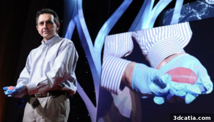
The conceptual roots of 3D bioprinting trace back to 1988, when Dr. Robert J. Klebe, a pioneering researcher at the University of Texas, introduced his “cytoscribing” process. This innovative method involved the micropositioning of cells using a modified inkjet printer to construct both two and three-dimensional synthetic tissues. This early work laid a foundational understanding for how conventional printing techniques could be adapted for biological applications. A significant leap forward occurred in 2002 when Professor Anthony Atala of Wake Forest University successfully created the first organ using bioprinting techniques: a small-scale kidney. This achievement, though modest in size, demonstrated the tangible possibility of engineering complex organ structures.
Further fostering the development and commercialization of bioprinting, Organovo, the world’s first commercial bioprinting laboratory, was established in San Diego, California, in 2010. Collaborating with Invetech developers, Organovo swiftly embarked on creating one of the earliest commercially available bioprinters, the NovoGen MMX. Since its inception, Organovo has been instrumental in advancing bone tissue engineering, and notably, has made significant strides in producing functional liver tissues for transplant research. Their work exemplifies the transition of bioprinting from a purely academic pursuit to a field with immense commercial and therapeutic potential.
Professor Anthony Atala
A truly monumental breakthrough in the field occurred in 2019, when a team of researchers at Tel-Aviv University (TAU) successfully 3D printed a heart using human cells. This remarkable achievement was particularly significant because the bioprinted heart perfectly replicated the immunological, cellular, and anatomical properties of a human patient. Although the initial prototype was the size of a rabbit heart, its intricate cellular and vascular complexity marked an unprecedented milestone. Prof. Tal Dvir, who spearheaded the study, articulated the significance: “In the past, they have been able to 3D print the structure of a heart, but not with cells and blood vessels. Our results demonstrate the potential of our approach for making customized tissues and organ replacement in the future.” Following this pioneering work by the TAU team, BIOLIFE4D also made headlines by bioprinting a miniature human heart, becoming the first company in the United States to achieve this feat, further validating the rapid advancements in creating patient-specific organ models.
The momentum continued into 2022, with researchers at Boston University developing a 3D bioprinted device dubbed the “Miniaturized Precision-Enabled Unidirectional Microfluidic Pump,” or miniPUMP. The most astonishing characteristic of the miniPUMP is its ability to autonomously beat, much like a natural human heart, thanks to its integrated living tissue. This innovation represents a crucial step forward in cardiovascular research, providing an invaluable tool for studying the intricate mechanics of heart function and the progression of various heart diseases. Concurrently, numerous other small-scale organ reproduction projects have seen success in recent years, collectively indicating a robust and accelerated research trajectory toward the ultimate goal of printing full-sized, functional organs.
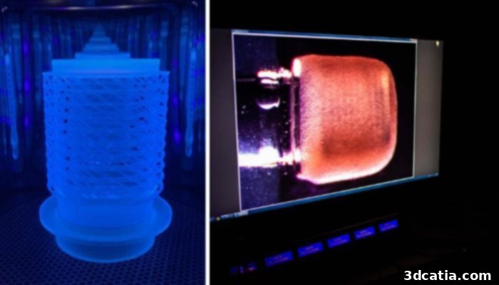
The miniPUMP was designed by researchers at Boston University (photo credits: Jackie Ricciardi)
Despite these remarkable advancements and the immense promise of 3D bioprinting, the technology is not without its significant challenges. A primary limitation often impeding its widespread development and adoption is the prohibitively high cost associated with research, specialized equipment, and premium biomaterials. Furthermore, profound ethical questions surrounding the creation and manipulation of living tissues and organs continue to emerge and demand careful consideration. Nevertheless, the field is dynamic, with continuous innovation leading to the development of new, more cost-effective, and sophisticated bioprinting techniques that are steadily gaining traction and pushing the boundaries of what is possible.
Understanding the 3D Bioprinting Process and Techniques
3D bioprinting offers an extensive range of applications within medicine and biomedical research. At its core, the underlying process for nearly every application involves a systematic approach: data collection, rigorous processing, and subsequent transformation of this data into a precise digital design. For instance, in the development of functional tissues and implants that accurately mimic human morphological and architectural characteristics, the process typically begins with detailed patient-specific anatomical data.
In simplified terms, the multi-stage bioprinting process encompasses:
- **Image and Cell Acquisition:** Obtaining high-resolution diagnostic images (such as CT scans, MRI scans) and harvesting specific tissues and cells from the patient. This provides the blueprint and the biological building blocks for the print.
- **Digital Reconstruction and Design:** Utilizing advanced bioengineering software and sophisticated techniques to reconstruct or design the damaged tissues or the specific parts to be reproduced. This stage translates biological data into a printable 3D model.
- **Biomaterial Selection:** Carefully selecting the most appropriate biomaterial or combination of biomaterials that will serve as the scaffold or matrix for cell growth. Factors like biocompatibility, mechanical properties, and degradation rate are crucial.
- **Bioink Preparation:** Incorporating the harvested cells into the chosen biomaterial to create a “bioink” – the printable living material. This step ensures cells are viable and uniformly distributed within the printing medium.
- **Printing Technology Selection:** Choosing the most suitable bioprinting technology based on the desired resolution, cell type, bioink viscosity, and the ultimate application of the printed construct.
Regarding specific bioprinting technologies, three primary methods are widely recognized and employed by the international scientific community: Inkjet Bioprinting, Extrusion Bioprinting, and Laser-based Bioprinting. Beyond these established techniques, a host of other more experimental and emerging methods are continuously being explored and developed, some of which will be highlighted subsequently.
Inkjet 3D Bioprinting: Precision Droplet Deposition
Inkjet 3D Bioprinting is one of the most established and widely recognized bioprinting technologies, fundamentally mirroring the operation of traditional inkjet printers. A key advantage of this method is the remarkable possibility of modifying conventional inkjet printers to function as sophisticated 3D bioprinters capable of handling bioinks. The process involves the precise deposition of discrete droplets of bioink (a solution containing cells and biomaterials) layer by layer onto a suitable substrate, such as a hydrogel or a Petri dish. The mechanism for ejecting these bioink droplets from the nozzle typically relies on one of two methods: thermal or piezoelectric actuation.
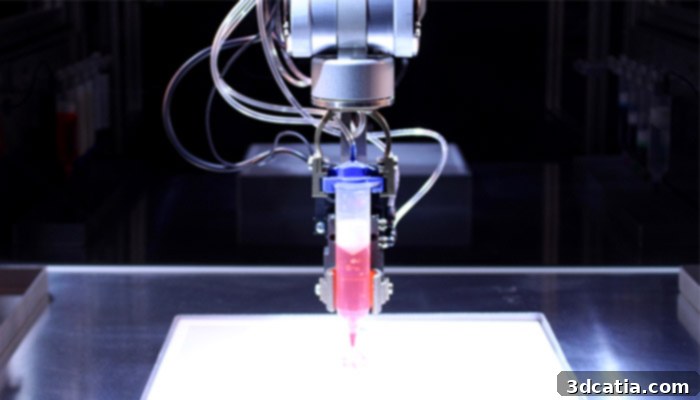
In the thermal inkjet bioprinting process, a localized heating element rapidly heats the bioink within the nozzle, creating a transient air bubble. The subsequent collapse of this bubble generates the necessary pressure to forcibly eject a precise droplet of bioink. Conversely, the piezoelectric method incorporates a piezoelectric ceramic crystal within the nozzle. This material possesses the unique property of expanding and contracting in response to an applied electric current. By carefully controlling small electrical pulses, the crystal’s rapid contraction or dilation expels the bioink. While inkjet bioprinting offers high resolution and speed, a notable disadvantage, particularly with the thermal method, is the potential for cell membrane damage due to heat or shear stress, which can lead to reduced cell viability and thus result in bioinks with poor cell content.
Despite these challenges, scientists have made significant advancements in utilizing inkjet bioprinting to understand the intricate patterns of molecules, cells, and even rudimentary organoids. This capability to precisely replicate cellular architectures facilitates the study of complex diseases like cancer and the development of potential targeted treatments. For instance, cells engineered to combat breast cancer have been successfully printed using inkjet bioprinting, retaining their functional integrity. This demonstrates promising prospects for creating viable living tissue structures and even functional organs in the future.
Organovo, a leader in the field, actively employs inkjet printing to develop sophisticated human models of inflammatory bowel disease (IBD). Through these meticulously bioprinted models, the company aims to gain deeper insights into the disease mechanisms, ultimately facilitating the discovery and development of superior drug therapies for debilitating conditions like ulcerative colitis and Crohn’s disease. Their 3D bioprinted tissues faithfully mimic crucial aspects of human biology and disease pathophysiology, extending their efforts to the successful reproduction of human liver tissues, which serves as a vital platform for drug toxicity screening.
Extrusion Bioprinting: Versatility and Scalability
Extrusion bioprinting has gained significant popularity due to its relative simplicity of execution, cost-effectiveness, and versatility. In this method, a solution comprising biomaterials and patient-specific cells, referred to as bioink, is continuously extruded through a nozzle. The extrusion force can be generated through various mechanisms, including pneumatics (air pressure), pistons, or screw-driven systems. As the printhead moves, it deposits continuous filaments of bioink, building up the desired model or tissue layer by layer. One of the notable advantages of this technology is its ability to operate effectively at room temperature, which is crucial for maintaining cell viability. Moreover, cells can be directly and homogeneously incorporated into the bioink mixture prior to printing, ensuring even distribution throughout the printed construct. Many popular bioprinters on the market, such as the Bioplotter and systems from EnvisionTec, leverage extrusion techniques, signifying its evolution from earlier inkjet bioprinting methods by offering greater control over material viscosity and cellular density.
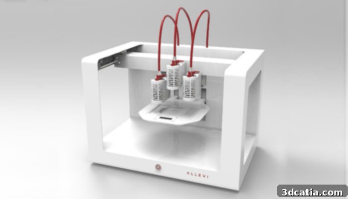
Allevi 3D Printer (photo credits: Allevi)
Laser-Assisted Bioprinting: High Precision and Contactless Deposition
Laser-assisted bioprinting, often employing a technique known as Laser-Induced Forward Transfer (LIFT), utilizes a precisely focused laser beam as its energy source to deposit biomaterials. This sophisticated technique typically involves three main components: a high-energy laser source, a “donor tape” (a transparent support coated with a layer of biological material), and a “receptor substrate.” The laser pulses are directed through the transparent side of the donor tape, impacting the biological material layer. This interaction causes rapid evaporation or ablation of a small portion of the material, generating a pressure wave that propels liquid biological material as droplets towards the receptor. These meticulously controlled droplets, containing cells encapsulated within a biopolymer, adhere to the substrate and initiate cellular growth and integration. A significant advantage of laser-assisted bioprinting is its nozzle-free and contactless nature, which minimizes mechanical stress on delicate cells. This allows for exceptionally high-resolution cell printing and precise droplet control of bioink. However, the advanced optical and laser systems required make this technology inherently expensive, with prohibitive costs limiting its widespread accessibility and deployment across many research and clinical settings.
Poietis, a pioneering French company in the bioprinting sector, specializes in designing and manufacturing advanced laser-assisted bioprinting machines. The company garnered significant attention through its high-profile partnership with L’Oréal, where they explored the ambitious goal of recreating hair follicles to stimulate hair growth. Furthermore, Poietis successfully developed Poieskin, an innovative human skin model meticulously constructed entirely through 3D bioprinting techniques. To provide a clearer understanding of the intricate laser-assisted bioprinting process, Poietis has produced an illustrative video demonstrating its operational principles and capabilities.
Emerging Techniques and Innovations in 3D Bioprinting
Stereolithography for Bioprinting: Light-Activated Tissue Fabrication
Stereolithography (SLA), a foundational additive manufacturing technique, is being adapted for bioprinting. This process involves the selective solidification of a photopolymer resin using focused ultraviolet (UV) light. For bioprinting applications, SLA is particularly well-suited because it can precisely print with hydrogels that are specifically engineered to be photosensitive. SLA offers unparalleled manufacturing accuracy and resolution, capable of creating intricate structures with fine details. However, its application in bioprinting is still under active development due to certain limitations. These include concerns regarding the biocompatibility and biodegradability of some polymers, potential adverse effects of UV light exposure on living cells, and the complexities associated with removing support structures without damaging delicate biological constructs.
Bioprinting with Acoustic Waves: Contactless Cellular Manipulation
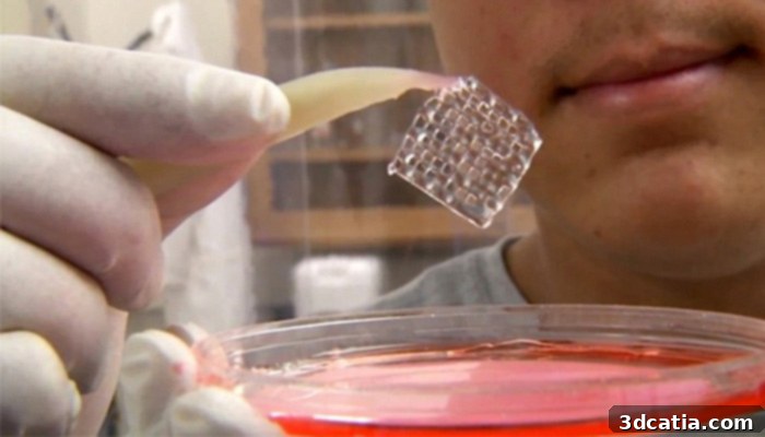
A revolutionary bioprinting technology has emerged from collaborative research at Carnegie Mellon University, Pennsylvania State University, and MIT, utilizing acoustic waves for precise cellular manipulation. This technique employs ‘acoustic clamps’ within a microfluidic device, enabling researchers to control the movement and arrangement of cells or microparticles through the application of superficial acoustic waves. By meticulously controlling the phase and frequency of these waves, researchers can precisely dictate where the waves converge along three spatial axes. At these specific convergence points, the acoustic waves generate a 3D trapping node capable of capturing and holding individual cells. These trapped cells are then systematically collected and arranged to form two-dimensional patterns, which can subsequently be stacked and assembled into three-dimensional biological constructs. This novel technique offers exceptional performance in terms of movement accuracy and, critically, provides a contactless method for cell handling, minimizing potential damage and preserving cell viability.
The rapid pace of innovation in bioprinting continues to yield new applications and techniques at a remarkable speed. Illustrative examples include the pioneering work at Northwest University in Illinois, where researchers successfully created a 3D printed ovary, offering new avenues for fertility research. Similarly, in Sweden, scientists have achieved the successful 3D bioprinting of human cartilage tissue, holding significant promise for regenerative therapies for joint injuries and degenerative conditions. While these medical advancements generate immense excitement and speculation about the future of healthcare, they also bring to the forefront a critical aspect that demands thorough exploration: the complex ethical implications inherent in such advanced biotechnologies.
The SWIFT Technique: Engineering Vascularized Tissues
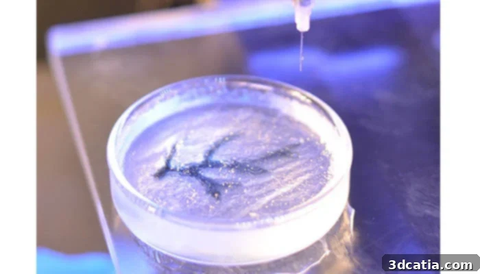
Researchers at Harvard’s Wyss Institute for Biologically Inspired Engineering have developed an ingenious bioprinting technique known as SWIFT (Sacrificial Writing Into Functional Tissue). As its name suggests, this innovative method focuses on bioprinting intricate vascular networks directly into living tissues. Specifically, SWIFT enables the 3D printing of functional vascular channels within living matrices composed of stem-cell-derived “organ building blocks” (OBBs). This approach is particularly transformative because it addresses one of the most significant hurdles in tissue engineering: the efficient vascularization of larger, thicker tissue constructs.
Rather than attempting the monumental task of 3D printing an entire organ cell by cell, SWIFT strategically concentrates on creating only the essential vascular infrastructure. This network is crucial for sustaining the viability of large quantities of OBBs within a tissue construct. These lab-grown, vascularized constructs, containing patient-specific cells, hold immense therapeutic potential for repairing and replacing damaged human organs. In a compelling experiment, organ-specific tissues were bioprinted with embedded vascular channels using the SWIFT technique and demonstrated sustained viability. In stark contrast, tissues grown without these crucial vascular networks experienced widespread cell death within a mere twelve hours, unequivocally highlighting the critical importance and effectiveness of vascularization for tissue survival and function.
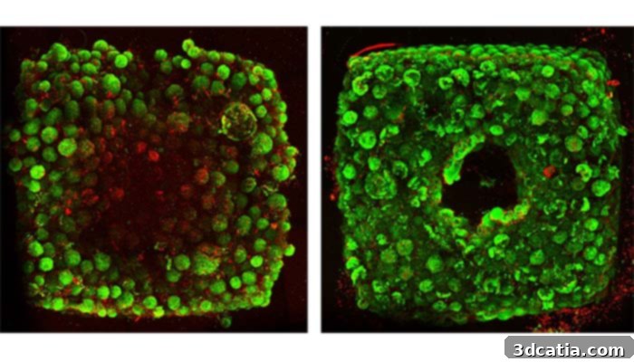
Organ-specific tissues printed with embedded vascular channels using SWIFT remained viable (right), while tissues grown without these channels experienced cell death within 12 hours (left).
The Future of 3D Bioprinting and the Era of Personalized Medicine
The medical community is increasingly embracing personalized medicine, a paradigm shift aimed at tailoring treatments to each patient’s unique genetic makeup, lifestyle, and specific disease characteristics. In this evolving landscape, 3D bioprinting plays a pivotal role, particularly in the in vitro reproduction of tissues for drug testing and the detailed study of specific diseases. This approach offers a significant advantage: it drastically reduces the reliance on animal testing and dramatically increases treatment effectiveness by allowing drugs to be tested on biological models that precisely reflect the individual characteristics of each patient. This level of specificity promises more targeted and efficacious therapies.
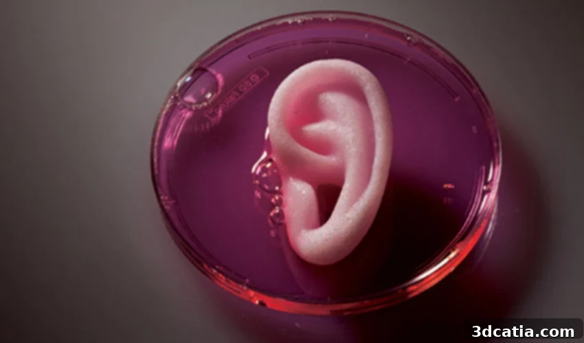
A compelling example of this personalized approach is the innovative work of Carcinotech, a company achieving remarkable success by 3D printing tumors derived directly from patient cells. These bioprinted tumor models serve as highly accurate platforms for testing various drugs, identifying higher-yielding therapies, and accelerating the development of new, more targeted, and combination cancer treatments. Beyond drug testing, the creation of implants or tissues based on patient cells has already transitioned from research to successful clinical applications. A prime illustration in this field is 3DBio Therapeutics, a company that designs and manufactures ear implants using collagen hydrogels infused with the patient’s own cartilage cells, offering bespoke regenerative solutions.
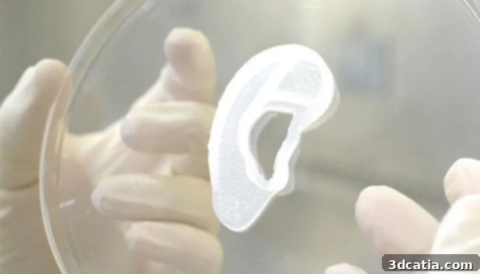
(Photo credits: 3DBio Therapeutics)
In terms of market trends, Grand View Research, a prominent market research firm, projects robust growth for the global bioprinting market, expecting it to reach an impressive $4.1 billion by 2026, with a Compound Annual Growth Rate (CAGR) of 19.5%. This growth is significantly driven by advancements in biomaterials and the synergistic integration of other cutting-edge technologies like Artificial Intelligence (AI). AI, for instance, is increasingly enabling scientists to quickly determine the optimal combination of biomaterials for converting cellular scaffolds into functional tissues. Bioprinting companies are anticipated to intensify their focus on developing a wider array of advanced biomaterials and enhancing bioprinting systems with multiple printheads, which will facilitate the simultaneous use of various bioinks within a single print. Furthermore, bioprinting software is expected to undergo significant upgrades, offering users more sophisticated control and expanded design options.
While the future of 3D bioprinting is exceptionally bright, it is imperative to address the main present and future concerns that accompany such transformative technology. Beyond the aforementioned high associated costs, a profound ethical debate looms concerning the broader consequences of personalized medicine and, critically, who will have equitable access to these life-altering advancements. Another significant ethical and practical challenge lies in rigorously testing the efficacy and long-term safety of these bioprinted treatments. Although analysis of various techniques suggests the plausible development of functional organs and tissues capable of replacing human ones, it remains exceedingly difficult to definitively assess whether a patient’s body will fully accept a new tissue or artificial organ without long-term clinical data. Moreover, robust regulatory frameworks and comprehensive legal guidelines must be meticulously prepared and established before these groundbreaking advancements can be made widely available to the general public, ensuring patient safety and societal confidence.
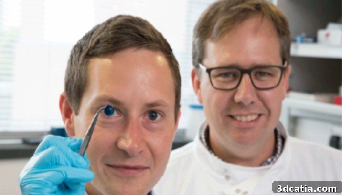
The creators of the first 3D printed cornea (photo credits: Newcastle University)
Furthermore, like any powerful new technology, 3D bioprinting carries the inherent risk of potential misuse, and it is no exception. If these technologies advance to the point of creating organs or tissues that are not merely replacements but are enhanced or “adaptable” based on specific human needs, we must deeply consider the ethical ramifications of such highly personalized medicine. This includes contemplating the potential creation of novel “superhuman” capabilities, such as bones engineered for exceptional resilience or lungs designed for significantly optimized oxygenation. These scenarios push the boundaries of medical ethics, necessitating proactive dialogue and robust oversight to ensure that such profound capabilities are developed and utilized responsibly for the betterment of humanity.
Do you believe that 3D bioprinting truly represents the future of personalized medicine? Share your thoughts and insights by leaving a comment below or connecting with us on our LinkedIn, Facebook, and Twitter pages! Don’t miss out on the latest advancements in 3D printing technology – remember to sign up for our free weekly newsletter here, delivered straight to your inbox! You can also explore all our informative videos on our dedicated YouTube channel.