
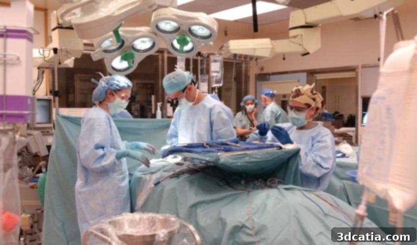
Revolutionizing Brain Cancer Treatment: 3D Bioprinting Active Glioblastoma Tumors for Personalized Medicine
In a groundbreaking advancement that promises to reshape the future of cancer treatment, researchers at Tel Aviv University have successfully utilized 3D bioprinting technology to create a fully active and viable glioblastoma tumor. Glioblastoma is notoriously known as one of the most aggressive and devastating forms of brain cancer, posing significant challenges for diagnosis, treatment, and prognosis. While the concept of creating a tumor might initially seem counter-intuitive, this innovative approach is hailed as a pivotal step forward in personalized medicine. By accurately replicating a patient’s specific tumor in a laboratory setting, scientists and medical professionals can now conduct comprehensive analyses and test various therapeutic strategies to identify the most effective treatment plan tailored to that individual patient. This pioneering 3D-bioprinted tumor model offers an unprecedented pathway for tackling brain cancers, which are frequently resistant to conventional treatments and are incredibly difficult to surgically remove due due to their invasive nature and critical location within the brain.
The application of 3D printing within the medical sector is not entirely novel, with established uses ranging from anatomical models for surgical planning to custom prostheses and orthoses. Furthermore, advanced bioprinting techniques have been instrumental in the development of engineered tissues and organs for regenerative medicine, such as the reconstruction of bones or complex organ structures. However, this new development from Tel Aviv University signifies a fundamental shift in how bioprinting is applied in the fight against diseases, particularly aggressive cancers like glioblastoma. Instead of focusing solely on structural replacement or generic drug screening, this research leverages 3D bioprinting to create living, functional replicas of tumors, complete with their intricate microenvironment. This enables doctors and researchers to gain deep insights into the specific characteristics of a patient’s cancer, allowing for highly customized treatment protocols. The ability to precisely recreate a tumor using bioprinting technology means that potential therapies can be rigorously tested outside the patient’s body, significantly improving the chances of determining the optimal and most effective course of action, thus ushering in a new era of truly personalized cancer therapy. This leap from general medical devices to active disease modeling is poised to dramatically accelerate therapeutic discovery and improve patient outcomes.
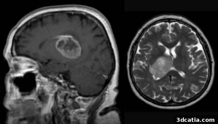
Glioblastoma tumors are known to be particularly aggressive as well as often difficult to treat
(Image Credits: Hellerhoff – Own work, CC BY-SA 3.0, https://commons.wikimedia.org/w/index.php?curid=18485437)
How is a 3D-Bioprinted Tumor Constructed for Advanced Cancer Research?
The pioneering study into the construction of these advanced tumor models was spearheaded by a distinguished team of researchers under the leadership of Professor Ronit Satchi-Fainaro. Professor Satchi-Fainaro holds several prominent positions at Tel Aviv University, including roles at the Sackler Faculty of Medicine, the Sagol School of Neuroscience, the Cancer Biology Research Center, the Nanomedicine Laboratory, and the Morris Kahn 3D-BioPrinting for Cancer Research Initiative. Her critical work was supported by a dedicated group of PhD students and researchers within her laboratory. The decision to pursue a 3D-bioprinted tumor model was a direct response to a significant challenge encountered in previous studies: a protein known as P-Selectin, which plays a crucial role in the spread and metastasis of cancer cells, could not be accurately reproduced or studied using conventional 2D plastic petri dish models. This fundamental inability to replicate complex biological interactions in standard laboratory settings is a major contributor to the alarmingly high failure rate of approximately 90% for new drugs during clinical trials. Recognizing this bottleneck, Professor Satchi-Fainaro’s team hypothesized that 3D bioprinting could provide a more physiologically relevant and reproducible platform, thereby rectifying the limitations of traditional research methods and paving the way for more successful drug development.
Indeed, their hypothesis proved correct. The researchers successfully replicated not only the glioblastoma cancer cells but also the crucial components of their intricate microenvironment. This includes surrounding cells, growth factors, and the extracellular matrix, all of which significantly influence tumor growth, aggression, and response to treatment. This comprehensive replication results in a far more accurate and biologically relevant model compared to traditional 2D cultures or even simplified animal models, leading to more reliable and predictive research outcomes. To achieve these sophisticated models, the research team carefully obtained tumor samples directly from glioblastoma patients. Utilizing these patient-derived cells and extracellular matrix components, the team ingeniously engineered a 3D-bioprinted tumor that incorporated a complex system of perfusable, blood vessel-like tubes. These finely structured channels allow for the precise flow of blood cells, nutrients, and, critically, anti-cancer drugs, mimicking the dynamics of a real tumor within the human brain. The success of this study was unequivocally demonstrated by the robust and reproducible predictions of optimal treatment responses for specific patients observed in the 3D-bioprinted models. This capability marks a monumental leap forward, offering a powerful tool for developing highly effective, patient-specific cancer therapies.
Professor Satchi-Fainaro articulated the immense potential of this innovative technology, stating: “If we take a sample from a patient’s tissue, together with its extracellular matrix, we can 3D-bioprint from this sample 100 tiny tumors and test many different drugs in various combinations to discover the optimal treatment for this specific tumor. This unparalleled ability to rapidly screen multiple therapies in parallel offers a truly personalized approach to cancer care, significantly reducing the guesswork and improving treatment efficacy. Alternately, we can test numerous novel compounds on a 3D-bioprinted tumor and decide which is most promising for further development and investment as a potential drug. This greatly streamlines the drug discovery pipeline, allowing for more efficient identification of effective therapeutic agents and minimizing the resources wasted on less promising candidates. But perhaps the most exciting aspect is finding novel druggable target proteins and genes in cancer cells – a very difficult task when the tumor is inside the brain of a human patient or model animal. Our innovation gives us unprecedented access, with no time limits, to 3D tumors mimicking better the clinical scenario, enabling optimal investigation.” This unprecedented platform effectively bridges the gap between basic research and clinical application, providing an invaluable tool for both immediate patient benefit and long-term drug development. For those interested in delving deeper into the specifics of this groundbreaking research, the full published study can be accessed HERE, or an explanatory video is available for a more visual understanding of the process and its implications below.
This development truly heralds a new era in oncology, offering hope for patients suffering from aggressive cancers like glioblastoma. By creating patient-specific tumor models, researchers can move away from one-size-fits-all treatments towards highly tailored therapies that are more likely to succeed. This not only promises to enhance individual patient outcomes but also has the potential to drastically reduce the time and cost associated with drug development, as preclinical testing becomes significantly more accurate and predictive. The ability to identify novel therapeutic targets directly within a human-like tumor environment without the complexities of living organisms is a game-changer for understanding cancer biology and accelerating drug discovery. This innovative bioprinting technology stands as a testament to the transformative power of advanced manufacturing techniques when applied to complex biological challenges, paving the way for smarter, more effective cancer interventions in the coming years. It represents a critical step towards personalized precision medicine, where each patient’s unique cancer is treated with a strategy designed just for them, maximizing efficacy and minimizing adverse effects.
What are your thoughts on this revolutionary 3D-bioprinted tumor technology? Do you believe it will be a pivotal tool for future cancer research and personalized treatment strategies? We invite you to share your insights and opinions in a comment below or engage with our community on our Facebook and Twitter pages. Don’t forget to sign up for our complimentary weekly newsletter to stay informed with all the latest news, breakthroughs, and advancements in the fascinating world of 3D printing, delivered directly to your inbox!
*Thumbnail Photo Credits: NIH Clinical Center via flickr