

Revolutionizing Regenerative Medicine: Terasaki Institute’s Breakthrough in 3D Bioprinted Muscle Tissue
The landscape of modern medicine is undergoing a profound transformation, largely driven by the rapid advancements in additive manufacturing. This innovative field, often synonymously referred to as 3D printing, has transcended its origins in rapid prototyping to become a cornerstone of various professional sectors, from automotive and aerospace to consumer goods. However, its most profound impact is arguably being felt in the medical sector, where 3D printing is not just improving processes but actively creating innovations that have the potential to save lives and drastically enhance patient care. Among these groundbreaking developments, researchers at the esteemed Terasaki Institute for Biomedical Innovation have achieved a monumental feat: the successful development and initial trials of functional 3D printed muscle tissue. This breakthrough represents a significant leap forward in regenerative medicine, offering unprecedented hope for patients suffering from muscle loss.
Historically, the primary method for addressing lost or severely damaged muscle tissue has been autologous transfer. This intricate surgical procedure involves harvesting healthy muscle mass from one part of the patient’s body and transplanting it to the affected area. While often effective, this approach is far from ideal. It introduces a secondary wound site, increasing the risk of complications such as infection, pain, and scarring. Furthermore, the removal of muscle from a healthy area can lead to a functional deficit at the donor site, creating a new problem while attempting to solve another. These inherent limitations underscore the critical need for alternative, less invasive, and more effective solutions for muscle regeneration. It is this pressing need that the researchers at the Terasaki Institute sought to address with their pioneering work.
In response to the challenges posed by conventional muscle tissue replacement, the Terasaki Institute team has engineered an innovative bioink designed to meticulously mimic the natural process of muscle formation. This sophisticated bioink is the cornerstone of their strategy to produce synthetic muscle tissue that is not only structurally sound but also functionally viable. This innovative bioprinting technology holds immense promise as a revolutionary solution for individuals who have lost muscle mass due to traumatic injuries, debilitating diseases, or necessary surgical resections. By providing a customizable and potentially limitless source of functional muscle, this research could fundamentally transform reconstructive surgery and rehabilitation, offering a path to recovery that was previously unimaginable.
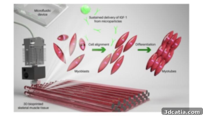
The bioink consists of a hydrogel, myoblast cells and microparticles (photo credits: Terasaki Institute)
The Science Behind the Bioink: Engineering Functional Muscle Tissue
Creating functional muscle tissue in vitro is an exceptionally complex endeavor. Skeletal muscle is a highly organized structure composed of various cell types, precisely regulated by intricate biochemical and biomechanical signaling pathways. To replicate this natural complexity, the Terasaki Institute researchers focused their efforts on integrating key biological components and growth factors into their bioink. A central element of their strategy was the utilization of insulin-like growth factor-1 (IGF-1), a potent hormone structurally similar to insulin that plays a critical role in the normal development and maintenance of bone and various tissues, including muscle. The controlled delivery of IGF-1 was paramount for guiding the differentiation and maturation of muscle cells.
The carefully formulated bioink developed by the team is a sophisticated composite of three primary substances, each playing a vital role in the success of the bioprinting process. First, a gelatin-based hydrogel provides the essential structural scaffold. Hydrogels, with their high water content and biocompatibility, closely mimic the extracellular matrix (ECM) of natural tissues, offering a supportive and nourishing environment for cellular growth and differentiation. Second, myoblast cells – the progenitor cells that eventually fuse to form muscle fibers – are suspended within this hydrogel. These cells are the building blocks of the new muscle tissue, programmed to differentiate and mature into functional muscle cells. Third, and critically innovative, are polylactic acid (PLGA) microparticles. These biodegradable microparticles are meticulously coated with IGF-1, acting as tiny reservoirs that continuously and steadily release the growth factor over time. This sustained release mechanism ensures that the myoblast cells receive the necessary biochemical cues for prolonged periods, promoting optimal muscle development and preventing the rapid degradation of IGF-1 that would occur if it were simply added to the culture medium.
From Lab to Life: Demonstrating Viability and Functionality
The efficacy of the Terasaki Institute’s bioink and bioprinting protocol was rigorously tested in the laboratory. A mere three days after the 3D printing process, a crucial milestone was achieved: the myoblast cells embedded within the hydrogel were confirmed to be highly viable, indicating that the printing process itself did not compromise their integrity or survival. This initial success paved the way for the subsequent and most impressive phase of the experiment. Over the course of ten days, these viable myoblast cells underwent successful differentiation and maturation, culminating in the development of complete synthetic muscle tissue. What was particularly remarkable was the observation that this cultured tissue began to exhibit spontaneous movements, a clear indicator of functional muscle fiber formation and contraction, closely mimicking the behavior of real, living muscle.
To further validate the potential of this groundbreaking technology, the cultured 3D printed muscle tissue was subsequently implanted into mice. This in vivo testing phase is critical for assessing how the engineered tissue integrates within a living biological system and whether it can maintain its viability and functionality outside of a controlled laboratory environment. The results were highly encouraging: researchers were able to confirm that the synthetic muscle tissue was not only successfully accepted by the host animal’s body, avoiding rejection, but also seamlessly combined and integrated with the existing muscle mass. This successful integration represents a monumental step, suggesting that the bioprinted tissue can establish the necessary connections and function synergistically with native tissues, a prerequisite for any clinical application.
The Future of Medicine: Implications and Outlook for 3D Printed Muscle Tissue
The development of 3D printed muscle tissue by the Terasaki Institute signifies a monumental advance in the field of regenerative medicine. By effectively mimicking the natural developmental processes of muscle formation, this sophisticated bioprinting technology is poised to revolutionize how we approach the replacement of lost or damaged muscle mass. The implications for patients are profound, offering new hope for individuals suffering from conditions ranging from severe traumatic injuries, such as those sustained in accidents, to debilitating degenerative diseases like muscular dystrophy, and even complex reconstructions following cancer surgery. Imagine a future where extensive muscle loss, which currently leads to chronic disability and reduced quality of life, could be effectively remedied with custom-printed, functional biological tissue.
While the initial results are incredibly promising, the path from laboratory breakthrough to widespread clinical application is often long and arduous. Currently, further extensive clinical studies and rigorous testing will be absolutely necessary. These critical steps will focus on meticulously confirming the long-term safety, durability, and efficacy of this process in complex biological systems, ultimately leading to its safe translation for treating human patients. Researchers will need to address challenges such as achieving vascularization – the formation of blood vessels within the printed tissue to supply nutrients and oxygen – and ensuring optimal nerve integration for controlled movement. However, despite these inherent challenges, the outlook for 3D printing muscle tissue remains overwhelmingly promising for the future of medicine. This innovative approach heralds a new era of personalized medicine, where custom-engineered tissues can restore function, improve recovery, and significantly enhance the lives of countless individuals globally. For those eager to delve deeper into the specifics of this pioneering research, additional detailed information is available by clicking HERE.
What are your thoughts on the groundbreaking potential of 3D printed muscle tissue and its impact on regenerative medicine? We invite you to share your insights and comments below, or engage with our community on our LinkedIn, Facebook, and Twitter pages! Stay informed about the very latest advancements in 3D printing technology by signing up for our free weekly Newsletter here, delivered directly to your inbox. You can also explore all our informative videos and content on our dedicated YouTube channel for more visual insights into the world of additive manufacturing.
*Cover Photo Credits: de Barros et al./Terasaki Institute for Biomedical Innovation