
Revolutionizing Medicine: Exploring 3D Bioprinting, Nanotechnology, and Advanced Tissue Engineering at TU Delft
The fields of 3D printing and bioprinting are rapidly transforming medical research, offering unprecedented opportunities for understanding complex biological systems and developing innovative therapies. These cutting-edge technologies enable the creation of intricate 3D microstructures and nanostructures that precisely mimic the natural environment of human tissue, providing a crucial platform for studying cellular behavior in a physiologically relevant context. Specifically, 3D bioprinting stands out as an invaluable tool for replicating damaged tissues or tumors using human cells, allowing researchers to accurately identify their functions and develop highly effective, personalized treatment strategies. This capability holds immense promise for the future of medicine, moving us closer to therapies tailored to individual patient needs and reducing the reliance on traditional, often less accurate, experimental models.
Among the leading institutions pioneering research in the application of 3D printing techniques for nanotechnology in the biomedical sector is Delft University of Technology (TU Delft). This esteemed university is at the forefront of leveraging advanced manufacturing to tackle some of the most pressing challenges in healthcare. To gain deeper insights into the groundbreaking projects undertaken by the university and the intricacies of working in this specialized domain, particularly in the fabrication of advanced 3D microstructures, we had the privilege of speaking with Angelo Accardo. As an Associate Professor in the Department of Precision and Microsystems Engineering at TU Delft, Professor Accardo is a driving force behind these advancements, pushing the boundaries of biomedical engineering and regenerative medicine through innovative research.
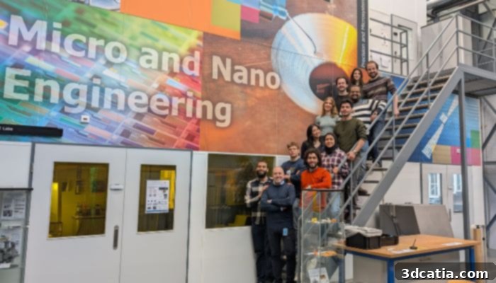
The dedicated TU Delft team with Professor Angelo Accardo, advancing 3D bioprinting and nanotechnology for future medical breakthroughs.
Meet Professor Angelo Accardo: A Leader in Biomedical Nanotechnology and Precision Engineering
Professor Angelo Accardo introduces himself as an Associate Professor within the prestigious Department of Precision and Microsystems Engineering at Delft University of Technology (TU Delft) in the Netherlands. His work spans multiple critical disciplines, focusing on the sophisticated intersection of engineering principles, advanced materials science, and fundamental cell biology to address significant medical challenges. His expertise lies in developing highly precise micro- and nanoscale devices and structures that can interact with biological systems in unprecedented ways.
Professor Accardo’s Pioneering Journey into Additive Manufacturing for Biomedical Applications
Professor Accardo’s foundational engagement with additive manufacturing techniques began during his post-doctoral research tenure at the LAAS-CNRS laboratory in Toulouse, France. It was within this innovative environment that he embarked on exploring the profound potential of light- and laser-assisted additive manufacturing technologies. His early and instrumental work focused particularly on stereolithography and two-photon lithography. These sophisticated fabrication methods, known for their ability to create incredibly intricate and precise 3D structures, laid the essential groundwork for his subsequent pioneering research. This initial exposure ignited his passion for utilizing these advanced techniques to generate bespoke micro- and nanostructures that could intimately interact with biological systems, ultimately driving advancements in the biomedical field.
The Extraordinary Promise and Enduring Challenges of 3D Printing in Nanotechnology and Biomedical Engineering
When discussing the intricate applications of 3D printing in nanotechnology and the biomedical sector, Professor Accardo highlights both the extraordinary benefits and the significant, ongoing challenges. These advanced fabrication techniques, particularly two-photon lithography, offer an unparalleled ability to develop micro- and nanostructures with exceptionally high levels of resolution, often reaching down to an astonishing 100 nanometers. This exquisite precision is not just an engineering feat; it is vital because at such minute scales, researchers can meticulously engineer micro-scaffolds that effectively interface biomimically with healthy or diseased cells. This biomimicry ensures that the engineered environment closely resembles the natural biological context, providing a far more accurate and relevant platform for cellular studies than traditional, two-dimensional culture methods.
Such precisely engineered microstructures are therefore indispensable for understanding how cells behave, grow, and function within an environment that closely mirrors natural human tissue. The invaluable knowledge gained from these detailed studies is a critical stepping stone towards the next transformative application: employing these sophisticated 3D microstructures in the burgeoning fields of tissue engineering and regenerative medicine. This involves the ambitious yet attainable goal of regenerating damaged tissues, paving the way for revolutionary treatments for severe injuries, chronic diseases, and even organ failure, thereby enhancing patient recovery and quality of life.
Despite these immense benefits, several significant challenges persist in this rapidly evolving field. One of the foremost hurdles is ensuring that these biomaterials are completely biocompatible. This characteristic is paramount to prevent any adverse reactions, immunological responses, or inflammation from the surrounding native tissue once the engineered structure is implanted into the body. Furthermore, these materials must also be biodegradable, meaning they should naturally “vanish” or be absorbed by the body in a controlled manner once the regeneration of the targeted tissue is successfully achieved. This requires careful consideration of material properties, degradation rates, and mechanical integrity over time, especially when designing and employing scaffolds with relatively large overall dimensions, sometimes up to several centimeters, for macroscopic tissue repair and integration.
Another critical challenge lies in the current inherent limitations of two-photon fabrication technology. While undeniably accurate and capable of producing intricate designs, it is still relatively “slow” when compared to the demands of large-scale production or rapid prototyping required for clinical translation. To overcome this throughput bottleneck, Professor Accardo emphasizes the urgent need to explore novel approaches that can significantly increase fabrication speed. Promising avenues include the development of two-photon “grayscale” lithography, which allows for varying laser intensity to create different features simultaneously, thereby enhancing efficiency, and the implementation of two-photon multibeam systems, which enable parallel fabrication over a wider area. Advancements in these areas are essential to scale up production, reduce manufacturing costs, and make these sophisticated techniques more viable for widespread biomedical applications.
Cutting-Edge Research on 3D Microstructures: Advancing Mechanobiology, Disease Models, and Tissue Engineering at TU Delft
Professor Accardo’s current research at TU Delft is meticulously centered on the innovative design and fabrication of 3D microstructures, targeting three distinct yet deeply interconnected applications: mechanobiology, in-vitro models of disease, and tissue engineering. His team’s groundbreaking work is consistently pushing the boundaries of what is scientifically and technologically possible in understanding and manipulating biological systems at their most fundamental levels.
Mechanobiology: Deciphering Cellular Response to Physical Cues with Nanostructures
One significant area of his impactful research, recently featured on the cover of the prestigious journal Advanced Functional Materials, delves into the fascinating field of mechanobiology. This particular study focuses on innovative 3D nanostructures fabricated using two-photon lithography, meticulously designed to have diameters of just a few hundred nanometers. These precise dimensions are highly significant because they closely resemble the natural fibers of the brain’s extracellular matrix (ECM) – the intricate, supportive network that surrounds and influences brain cells – as well as the delicate structures of filopodia. Filopodia are small, dynamic, finger-like protrusions that cells actively use to probe their immediate surroundings, sense their environment, and establish crucial interactions with other cells and their substratum.
A key innovation central to this research is the remarkable ability to adjust the aspect ratio of these nanostructures. This precise control allows the engineered structures to provide a relatively low effective shear modulus. This specific property refers to the elastic modulus, or perceived stiffness, detected by cells as they crawl over and interact with the nanostructures. By carefully tuning this mechanical characteristic, Professor Accardo and his team can create biomimetic environments that accurately approximate the intrinsic softness and mechanical properties of natural human brain tissue. This sophisticated mimicry is profoundly important because these precise topographical and mechanical signals exert a significant influence on the growth, development, and essential directionality of neuronal networks. Understanding these intricate interactions is absolutely critical for advancing our fundamental knowledge of brain development, function, and the progression of neurological diseases.
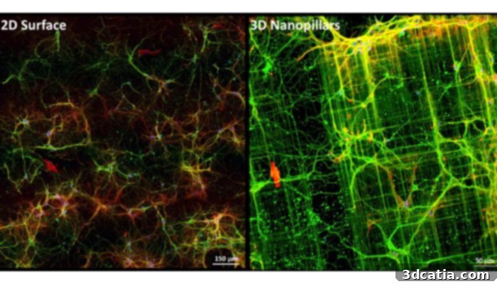
A compelling visual comparison illustrating the profound difference in neuronal growth and complexity in 2D (left) versus highly biomimetic 3D environments (right), enabled by advanced 3D microstructures. The 3D model allows for more natural cell morphology and network formation.
This innovative 3D bioprinting approach offers substantial advantages over conventional “Petri dish” methods, which have long been the standard in cell biology. In traditional setups, cells are typically cultured on flat, overly rigid plastic or glass surfaces, which are vastly dissimilar from the complex, dynamic, and soft mechanical properties of natural tissues within the body. Professor Accardo’s method, by contrast, provides topographical and mechanical signals that are far more physiologically relevant. These nuanced signals profoundly influence crucial neuronal cell properties, such as the overall directionality and organization of the neuronal network and the intricate morphology of growth cones. Growth cones are dynamic, highly motile structures located at the tips of growing axons that guide neurons as they actively explore the surrounding extracellular matrix to form precise connections with other neurons – a process absolutely vital for proper brain wiring and function. Understanding and precisely controlling these intricate aspects are pivotal for both fundamental neurological research and the development of regenerative therapies.
In-Vitro Disease Models: Ushering in a New Era for Neurodegenerative Research
The healthy development of neuronal networks and the normal morphology of growth cones are often profoundly and deleteriously affected by debilitating neurodegenerative diseases such as Alzheimer’s disease (AD) and Parkinson’s disease (PD). Through their advanced 3D platform, Professor Accardo and his team have not only demonstrated the impressive capability to guide the formation of physiologically relevant neuronal networks but also to quantitatively characterize growth cone morphology with unprecedented precision. This level of detail and control allows for a significantly deeper understanding of how these devastating diseases disrupt fundamental neuronal processes at the cellular and network levels.
Professor Accardo envisions the strategic application of their sophisticated platform as an advanced in-vitro engineered disease model. This model could prove instrumental in elucidating the precise influence of major pathological hallmarks associated with Alzheimer’s disease, such as the accumulation of amyloid plaques and neurofibrillary tangles, and Parkinson’s disease, characterized by the presence of Lewy bodies. By accurately simulating these complex conditions in a controlled 3D environment that closely mimics the brain’s natural setting, researchers can gain critical insights into their specific impact on neuronal network directionality and growth cone development. This could potentially lead to the identification of novel therapeutic targets and pathways. Furthermore, this innovative approach dramatically reduces the reliance on traditional animal models, offering a more ethically sound, cost-effective, and often more predictive research pathway for drug discovery and disease understanding.
Combating Brain Cancer: Developing Advanced 3D Models for Proton Therapy Optimization
Beyond neurodegenerative diseases, Professor Accardo’s team has also made significant strides in developing sophisticated 3D models for the improved treatment of brain cancer, specifically glioblastoma. Glioblastoma is recognized as one of the most aggressive and challenging forms of brain tumor, known for its rapid progression and resistance to conventional therapies. This particular line of research is intensely focused on optimizing proton therapy, an advanced and highly precise radiation treatment technique. Unlike conventional X-rays used in traditional radiation therapy, which deposit energy along their entire path, proton therapy utilizes subatomic components – protons – to precisely target cancer cells. This inherent precision allows for minimal damage to surrounding healthy tissue, making it a highly desirable and safer treatment modality, especially for sensitive organs like the brain.
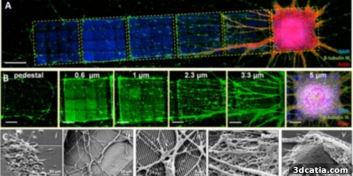
To achieve this critical goal, the team develops intricate three-dimensional structures that meticulously mimic the microvascular system of the brain – the complex network of tiny blood vessels and capillaries where glioblastoma cells typically grow, proliferate, and metastasize. These biomaterial-based 3D structures are ingeniously engineered to provide crucial mechanical, biochemical, and geometric stimuli to the glioblastoma cells. This close replication of the brain’s internal architecture, including the shapes of capillaries and blood vessels, creates a highly realistic and complex environment that glioblastoma cells encounter within the brain’s vasculature. This biomimicry is absolutely essential for establishing an accurate and predictive model to study tumor behavior, drug delivery, and crucially, treatment response to radiation.
Once the glioblastoma cells are meticulously cultured within this highly biomimetic 3D environment, they are carefully transported to the Holland Proton Therapy Center. Here, the cultured cells are precisely exposed to varying dosages of proton radiation. This unique and controlled in-vitro setup enables researchers to accurately calibrate the exact proton radiation dosages required to effectively damage the DNA of the cancer cells, leading to their programmed destruction, all within a laboratory setting. Crucially, this innovative approach allows for extensive experimentation and the optimization of proton therapy protocols with remarkable precision, entirely circumventing the ethical and logistical complexities traditionally associated with animal models. This accelerates the development of more effective, personalized, and safer treatments for patients battling glioblastoma, offering a beacon of hope in a challenging medical landscape.
The Path to Academia: Essential Qualifications and Skills for a Professor in Biomedical Engineering
Addressing the fundamental requirements for a highly specialized academic position such as his own, Professor Accardo emphasizes that a strong foundational education in engineering or physics is profoundly beneficial. Such a background helps cultivate a rigorous mindset well-suited to the scientific method, fostering critical thinking, logical reasoning, and sophisticated problem-solving skills – all essential attributes for conducting robust and impactful research. However, he quickly notes that academic pursuit in this rapidly evolving and inherently interdisciplinary field necessitates a commitment to continuous, lifelong learning. Over the years, one acquires a vast array of knowledge that extends far beyond initial technological aspects, deeply immersing oneself in crucial subjects like cell biology and neuroscience – disciplines absolutely critical for comprehensively understanding the intricate biological systems they aim to influence or repair.
Furthermore, to successfully hold a faculty position within a dynamic and competitive academic environment like the Netherlands, it is imperative to develop robust management and leadership skills. These skills are not merely about effectively overseeing one’s own research group, which typically involves guiding PhD students, postdocs, and technical staff, but also extend to fulfilling broader strategic roles within the university. This could encompass contributing significantly to curriculum development, actively participating in departmental committees, or leading collaborative research initiatives across different faculties or institutions. All these roles require a sophisticated blend of scientific acumen, administrative capability, and interpersonal leadership.
Wisdom for Aspiring Academics: Professor Accardo’s Advice for Future Professors
For individuals aspiring to embark on a challenging yet incredibly rewarding career as a professor, Professor Accardo offers invaluable advice centered around a core set of deeply ingrained personal attributes: “Passion, perseverance, and patience.” He stresses emphatically that these characteristics are not just desirable but are absolutely essential within the demanding and often unpredictable realms of academia and cutting-edge research. The academic journey frequently involves unforeseen setbacks, periods of intense effort and long hours, and the consistent need for sustained dedication over many years to achieve significant breakthroughs and recognition.
In addition to these foundational personal traits, Professor Accardo emphasizes the paramount importance of perpetually nurturing one’s curiosity. A successful professor must remain constantly inquisitive, always seeking new knowledge, fresh perspectives, and innovative solutions to complex problems. This continuous intellectual engagement is inextricably coupled with the necessity of staying acutely abreast of current scientific trends and emerging technological advancements. This vigilance is crucial not only for pushing the very boundaries of research but also for successfully writing compelling and competitive research projects that attract vital funding – an indispensable component of academic success and sustainability. Finally, Professor Accardo highlights the indispensable need for a strong predisposition toward teaching. A professor’s role extends significantly beyond personal research endeavors; it involves effectively passing on not only complex scientific notions but also an efficient modus operandi – a critical way of thinking, approaching problems, and engaging in scientific inquiry – to the next generation of engineers and scientists. This dedicated mentorship ensures the continuity, growth, and advancement of scientific progress for years to come.
What are your thoughts on Professor Angelo Accardo’s pioneering work on 3D microstructures, bioprinting, and advanced tissue engineering at TU Delft? We invite you to share your insights and comments below, or connect with us on our LinkedIn, Facebook, and Twitter pages! Don’t miss out on the latest advancements in additive manufacturing – remember to sign up for our free weekly Newsletter here, delivering the most relevant 3D printing news directly to your inbox! For more fascinating videos and discussions, visit our YouTube channel. Additionally, explore further medical and dental 3D printing news and articles HERE.