
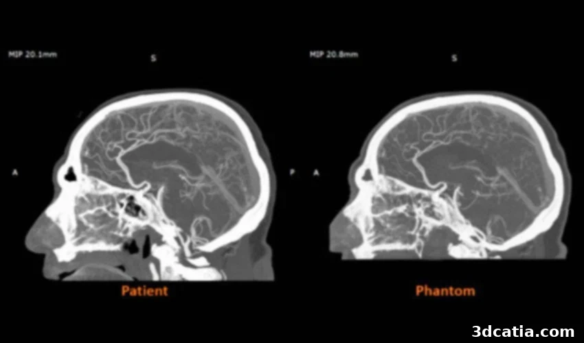
Stratasys RadioMatrix™: Revolutionizing Medical Imaging with Radiopaque 3D Printing
Stratasys has announced the full U.S. availability of RadioMatrix™, an innovative radiopaque 3D printing material designed to advance medical imaging education, device testing, and research. Previously accessible only to select programs in Europe, RadioMatrix™ is now available for healthcare providers, imaging centers, and medical device manufacturers across the United States, enabling them to seamlessly integrate this groundbreaking technology into their workflows.
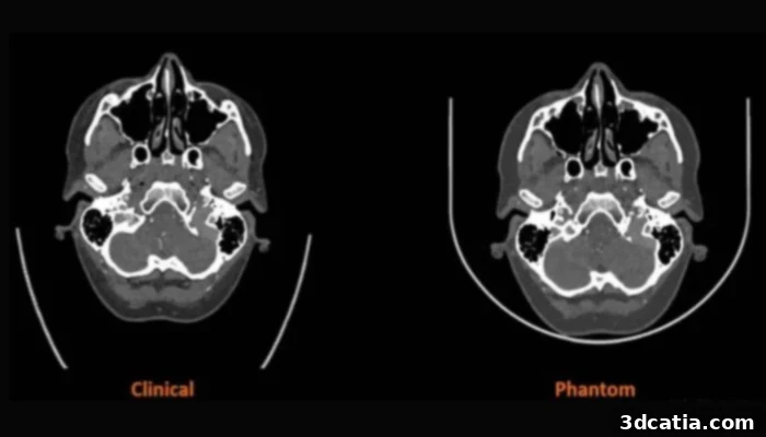
RadioMatrix™ distinguishes itself as the first and only 3D printing material that provides controlled radiopacity. This unique feature allows users to precisely match the appearance of specific tissues on CT and X-ray scans, achieving a high degree of accuracy. Compatible with the Stratasys J750 and J850 Digital Anatomy™ printers, which already excel at producing lifelike soft tissue and bone models, RadioMatrix™ enhances these models by replicating anatomical behavior under imaging conditions. This expansion of capabilities significantly increases the value of these models for teaching, research, and medical device development.
The integration of RadioMatrix™ into medical imaging promises to refine diagnostic accuracy and procedural training. By enabling the creation of anatomically precise and radiologically accurate models, Stratasys is pushing the boundaries of what’s possible in healthcare simulation and education.
Precise Control Over Radiopacity: A Game Changer in Medical Imaging
A critical advantage of RadioMatrix™ is its exceptional ability to fine-tune Hounsfield unit (HU) values. This precise tuning enables printed structures to closely mimic bone, gray matter, fat, veins, or any other anatomical feature expected in a clinical scan. Early collaborations between Stratasys and Siemens Healthineers have demonstrated that RadioMatrix™ phantoms can match human tissue densities with deviations as small as a single Hounsfield unit (HU). This unparalleled level of precision results in models that are nearly indistinguishable from real tissue on a CT scan, supporting the accurate replication of even the most complex anatomical structures.
The capacity to achieve such high fidelity makes RadioMatrix™ particularly well-suited for a broad range of applications, including advanced imaging education, detailed studies of pathology, and the rigorous validation of CT protocols and imaging algorithms. By providing realistic and customizable phantoms, RadioMatrix™ is enhancing the quality and effectiveness of medical research and training.
The implications of this technology extend beyond mere replication. The ability to create customized phantoms that accurately represent specific patient anatomies or pathological conditions allows for personalized medicine approaches in training and planning. Surgeons can practice complex procedures on models that mirror the unique challenges they will face in the operating room, leading to improved patient outcomes and reduced risks.
Benefits for Radiology Education: Redefining Training Standards
For radiology programs, the introduction of RadioMatrix™ represents a significant advancement. Traditional methods, such as the use of cadavers, are often costly, difficult to access, and limited in number. While traditional phantoms offer greater availability, they typically lack the realism and customization needed to effectively simulate specific pathologies. RadioMatrix™ provides a repeatable and scalable alternative, empowering educators to produce the exact anatomy or disease state they need to teach. These 3D printed models can be reused for consistent and repeatable training sessions across larger groups of students, ensuring that all trainees receive a standardized and high-quality learning experience.
Furthermore, RadioMatrix™ models can be easily updated and modified to reflect the latest advancements in medical knowledge and techniques. This adaptability is crucial in a rapidly evolving field like radiology, where new imaging modalities and diagnostic criteria are constantly emerging. By using RadioMatrix™, educators can ensure that their training programs remain current and relevant, preparing students to meet the challenges of modern medical practice.
“Providing full availability of RadioMatrix™ in the U.S. is a major step in providing cutting edge imaging education and training,” said Erez Ben Zvi, Vice President of Healthcare at Stratasys. “By giving radiologists and device manufacturers the ability to print ultra realistic, customized radiographically accurate models, we are helping replace traditional phantom solutions and reliance on cadavers with customizable, repeatable, and scalable alternatives.”
Growing Clinical and Research Applications: Expanding the Horizon of Medical Innovation
The versatility of RadioMatrix™ extends beyond education and training. In the United Kingdom, institutions like Beaumont Hospital and CPI have already leveraged the material to produce cerebral angiography models for training in imaging-guided procedures. These early successes demonstrate that printed radiopaque anatomy can support more consistent and controlled training environments, leading to improved proficiency and reduced error rates.
The material also offers significant benefits to medical device manufacturers and researchers who require reliable test models for the development and validation of new devices and imaging tools. Instead of relying on donor material or generic phantoms, teams can now print standardized structures tailored to their specific studies. This capability streamlines the research and development process, accelerating the pace of innovation in the medical field. With expanded access now available in the U.S., more institutions are expected to adopt radiopaque 3D printing and deepen their use of Digital Anatomy technology.
The standardization and reproducibility offered by RadioMatrix™ are particularly valuable in clinical trials. By using identical phantoms across multiple sites, researchers can minimize variability and ensure that their results are accurate and reliable. This level of control is essential for obtaining regulatory approval for new medical devices and therapies.
To learn more about the RadioMatrix™ materials, click HERE.
What are your thoughts on the increasing accessibility of radiopaque 3D printing for hospitals and imaging programs in the United States? How do you see this technology impacting the future of medical education and research? Share your perspectives in the comments below or on our LinkedIn and Facebook pages. Stay informed about the latest developments in 3D printing by subscribing to our free weekly Newsletter, delivering the news directly to your inbox. Explore our video content on our YouTube channel. For more medical and dental 3D printing news, visit our dedicated page here.
*All Photo & Video Credit: Stratasys