
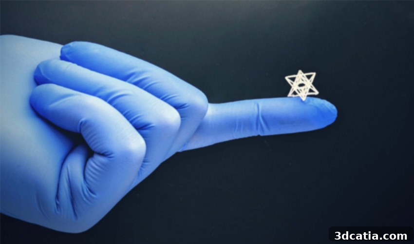
Revolutionizing Regenerative Medicine: RMIT’s NEST3D Bioprinting Method for Microscopic Medical Implants
The field of regenerative medicine is constantly pushing the boundaries of what’s possible, particularly in the creation of advanced medical implants designed to repair or replace damaged tissues and organs. At the forefront of this innovation, a groundbreaking collaboration between researchers at the Royal Melbourne Institute of Technology (RMIT University) and medical professionals from St Vincent’s Hospital in Melbourne has yielded a revolutionary approach to bioprinting medical implants. This innovative technique, known as Negative Embodied Sacrificial Template 3D (NEST3D), promises to overcome significant hurdles in tissue engineering, particularly concerning the fabrication of intricate, microscopic structures with exceptional precision and material versatility. Unlike conventional bioprinting methods that directly deposit cells and biomaterials to build scaffolds, NEST3D adopts an indirect strategy: it involves 3D printing sacrificial molds with incredibly detailed cavities, into which biocompatible materials are subsequently injected. Once the injected material has solidified, the mold is simply dissolved in water, leaving behind a perfectly formed, intricate bio-scaffold. This ingenious method is set to be compatible with a broad spectrum of materials, opening new avenues for creating minuscule yet complex structures critical for advanced medical applications.
The Bioprinting Landscape: Promises and Persistent Challenges
In tissue engineering, bioprinting has emerged as a transformative technology, enabling the precise design and fabrication of devices that actively promote the reconstruction and regeneration of bones, muscles, cartilage, and various soft tissues. Typically, 3D printed scaffolds are meticulously designed and then implanted into a patient’s body. These scaffolds serve as temporary structural frameworks, acting as biological blueprints and supportive matrices that encourage the patient’s own cells to adhere, proliferate, and differentiate, thereby facilitating the healing and regeneration of injuries which can range significantly in severity and complexity. This direct approach to scaffold fabrication has seen considerable success, but it also faces inherent limitations, especially when striving for the highest levels of resolution and structural intricacy.
One of the most significant obstacles in conventional bioprinting today revolves around the physical constraints of the printing process itself. The size and complexity of the structures that can be reliably produced are often limited by the nozzle diameter of the bioprinter or the resolution of light-based techniques. Designing and fabricating scaffolds in the micron range, which is often essential for mimicking natural tissue architecture and promoting effective cell infiltration and vascularization, remains a formidable challenge. Direct extrusion bioprinting, for instance, requires the bio-ink to flow through a nozzle, which inherently restricts the minimum feature size. This often results in scaffolds that, while functional, may lack the ultra-fine detail and porosity necessary for optimal biological integration and long-term success. Recognising these challenges, the RMIT team questioned: why not adopt a more indirect, yet potentially more precise, approach to scaffold fabrication?
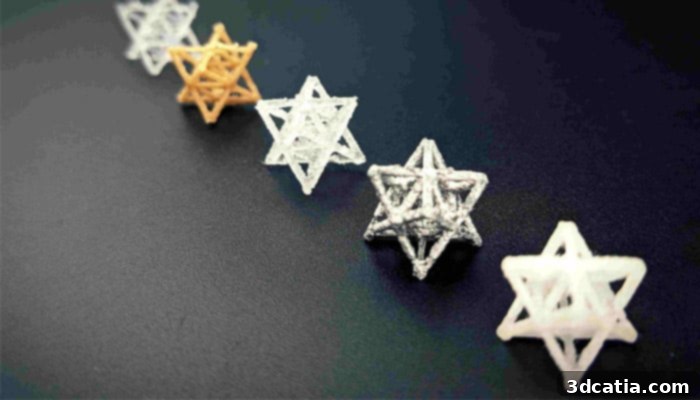
Using their method, researchers can design much more complex and smaller structures (photo credits: RMIT University)
Introducing NEST3D: An Indirect Approach to Precision Bioprinting
The brilliant solution conceived by this collaborative team is the NEST3D method, an acronym for Negative Embodied Sacrificial Template 3D. Instead of directly printing the scaffold, the researchers innovatively decided to 3D print a negative mold – essentially, a template with complex, intricately-patterned cavities that precisely define the intended shape of the future implant. This strategic inversion of the process allows for a level of detail and structural freedom previously difficult to achieve. The principle is elegant in its simplicity and profound in its implications.
The NEST3D Process Explained
The process begins with the 3D printing of the sacrificial mold. For this crucial step, the researchers found that a readily available and water-soluble material like PVA (polyvinyl alcohol) glue proved highly effective. PVA is excellent because it can be easily 3D printed using common FDM (Fused Deposition Modeling) printers, and crucially, it dissolves completely and cleanly in water without leaving any harmful residues. Once the intricate mold, complete with its complex cavities and channels, is printed and hardened, the next phase involves injecting biocompatible materials into these precisely formed holes. These injected materials are carefully chosen for their biological compatibility and mechanical properties, designed to create a welcoming and supportive scaffold for cells to thrive.
After the biocompatible material is injected and allowed to cure or harden within the mold’s cavities, the crucial dissolving step takes place. The entire assembly – the hardened mold with the embedded scaffold material – is immersed in water. The PVA glue, true to its nature, completely dissolves away. What remains is only the intricately shaped scaffold, perfectly formed to the negative image of the mold, now freed from its temporary casing. This method allows for the creation of structures that can be incredibly small, sometimes as tiny as a fingernail, yet possessing an unprecedented level of internal complexity and resolution. This indirect fabrication sidesteps the limitations of direct extrusion by using the mold’s internal features to define the scaffold’s minute architecture, rather than relying on the nozzle size of the bioprinter.
Unparalleled Versatility in Materials and Structures
One of the standout advantages of the NEST3D technique, as highlighted by Stephanie Doyle, a key researcher in this study, is its remarkable versatility. “The advantage of our advanced injection moulding technique is its versatility,” she explains. “We can produce dozens of trial bioscaffolds in a range of materials – from biodegradable polymers to hydrogels, silicones and ceramics – without the need for rigorous optimisation or specialist equipment. We’re able to produce 3D structures that can be just 200 microns across, the width of 4 human hairs, and with complexity that rivals that achievable by light-based fabrication techniques.” This statement underscores a critical breakthrough.
The ability to use such a wide variety of materials is paramount in regenerative medicine. Different tissues require different mechanical and biological properties. Biodegradable polymers, for instance, can provide temporary support and degrade naturally as new tissue forms. Hydrogels offer a soft, cell-friendly environment, mimicking the extracellular matrix. Silicones might be used for flexible implants, while ceramics are excellent for bone regeneration due to their stiffness and biocompatibility. The fact that NEST3D can accommodate these diverse materials without extensive re-calibration or highly specialized equipment makes it incredibly adaptable for various medical applications, from repairing cartilage in joints to regenerating nerve tissue or even supporting organoids for drug testing.
Furthermore, the precision achieved with NEST3D is truly exceptional. Structures just 200 microns (0.2 millimeters) across represent a significant leap in resolution for bioprinted implants. To put this into perspective, 200 microns is approximately the thickness of four human hairs. This micro-scale resolution is vital because natural tissues possess incredibly fine structures, and effective regeneration often requires scaffolds that can accurately replicate this microscopic complexity. By enabling the creation of such intricate and small structures, NEST3D opens the door for implants that can integrate more seamlessly with the body’s natural architecture, promoting better cell migration, nutrient supply, and ultimately, more successful tissue repair.
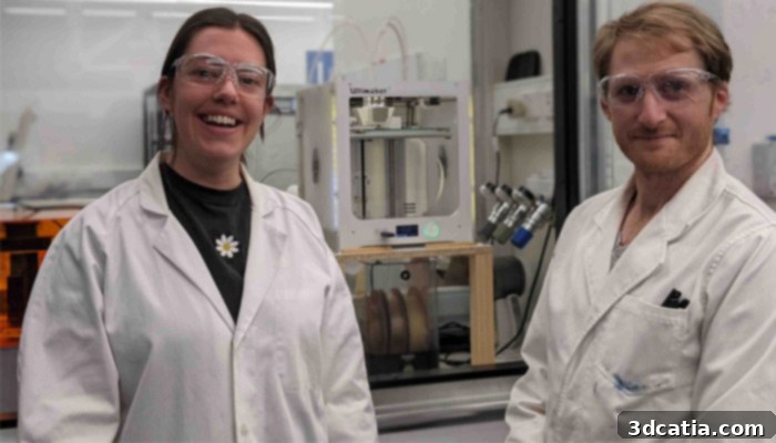
Stephanie Doyle and Dr. Cathal O’Connell in front of the 3D printer (photo credit: RMIT University)
Cost-Effectiveness and Accessibility: The FDM Advantage
Beyond its scientific elegance and technical prowess, the NEST3D method offers significant practical advantages in terms of cost-effectiveness, ease of use, and scalability. A major breakthrough is its compatibility with standard FDM (Fused Deposition Modeling) 3D printers – widely available, affordable machines used for basic plastic prototyping. This eliminates the need for expensive, specialized bioprinters that typically cost hundreds of thousands of dollars and require highly trained personnel to operate. By leveraging existing FDM technology, the barrier to entry for advanced bioprinting research and potential clinical application is dramatically lowered.
In traditional direct bioprinting, where biomaterials are extruded directly, the size of the machine’s nozzle is a critical limiting factor. The nozzle must be large enough to allow the bio-ink – which can often contain cells and viscous biomaterials – to flow through without clogging. This inherent physical constraint directly impacts the minimum feature size and overall resolution of the printed part, thereby limiting the complexity and fineness of the scaffolds that can be produced. However, with NEST3D, the FDM printer is used only to create the mold, which can be printed with much finer features and thinner walls than direct bioprinting allows. The critical difference is that the mold material (like PVA) is much easier to extrude through tiny nozzles than complex bio-inks. Once the fine mold is created, even the most delicate biocompatible materials can be carefully injected into these much thinner spaces, resulting in an exquisitely detailed final scaffold. This indirect approach effectively decouples the limitations of nozzle size from the resolution of the final implant, opening up a world of possibilities for microscopic and intricate designs.
Clinical Implications and Future Outlook
The immediate and future implications of the NEST3D method are vast. After rigorous testing, the researchers have confirmed that their produced scaffolds are both safe and non-toxic, a crucial prerequisite for any medical application. Their next objective is to accelerate cell reconstruction within these scaffolds by experimenting with different designs and material combinations. This ongoing research aims to optimize the scaffold’s architecture to best encourage cell growth, differentiation, and integration, ultimately paving the way for more effective tissue regeneration in patients.
For doctors and clinicians, this innovation represents a significant step towards more accessible and adaptable solutions for daily medical challenges. Professor Claudia Di Bella, an orthopedic surgeon at St. Vincent’s Hospital and a key collaborator in this project, eloquently emphasizes the importance of bridging the gap between clinical insight and engineering innovation. “A common problem faced by clinicians is the inability to access technological experimental solutions for the problems they face daily,” she states. “While a clinician is the best professional to recognize a problem and think about potential solutions, biomedical engineers can turn that idea into reality. Learning how to speak a common language across engineering and medicine is often an initial barrier, but once this is overcome, the possibilities are endless.” This sentiment perfectly encapsulates the spirit of the RMIT and St Vincent’s Hospital partnership, demonstrating how interdisciplinary collaboration can drive profound advancements in healthcare.
The NEST3D technique holds immense promise for various clinical applications, including but not limited to, reconstructive surgery for complex bone fractures, cartilage repair, nerve regeneration, and even the development of personalized drug delivery systems. The ability to rapidly prototype and produce highly customized, microscopically precise, and biologically compatible implants using cost-effective methods could revolutionize patient care. This groundbreaking research not only offers a powerful new tool for tissue engineering but also reinforces the critical role of synergistic collaboration between medical practitioners and engineering experts in shaping the future of regenerative medicine. Find more information on the university’s website HERE and in the video below:
Conclusion: A New Era for Bioprinted Medical Implants
The innovative NEST3D bioprinting method developed by RMIT University and St Vincent’s Hospital marks a significant leap forward in the quest for creating more effective and accessible medical implants. By transforming the traditional bioprinting paradigm from direct deposition to an indirect, mold-based approach, researchers have unlocked unprecedented levels of precision, structural complexity, and material versatility. This technique effectively sidesteps the common limitations of nozzle size in direct extrusion, allowing for the creation of intricate, microscopic scaffolds from a wide range of biocompatible materials. Its compatibility with readily available FDM 3D printers also makes it a remarkably cost-effective and scalable solution, democratizing access to advanced bioprinting capabilities. As the research progresses from safety testing to accelerating cell reconstruction, NEST3D stands poised to revolutionize regenerative medicine, offering clinicians and patients a promising pathway to enhanced tissue repair, personalized medical devices, and ultimately, improved quality of life. The future of bioprinted implants looks brighter and more accessible than ever before, thanks to this ingenious Australian innovation.
What do you think of this new research for bioprinting implants and its potential impact on healthcare? Let us know in a comment below or on ourFacebook,Twitter andLinkedIn pages! Sign up for our free weeklyNewsletter here, the latest 3D printing news straight to your inbox!