Applications
Using Cancer Cells And Computer Modeling, Researchers Have Optimized 3D Bioprinting
Researchers in a multi-university collaboration between the University of Waterloo, Canada, University of Swansea, and University of St Andrews, both UK, have conducted experiments on 3D bioprinted cancer cells over a period of 11 days, to compare to results from…
- 3 min read
- Applications
- Six more stories
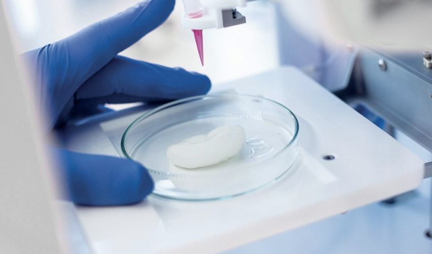
Researchers in a multi-university collaboration between the University of Waterloo, Canada, University of Swansea, and University of St Andrews, both UK, have conducted experiments on 3D bioprinted cancer cells over a period of 11 days, to compare to results from a cellular automata model. Their results showed that the model successfully simulated the bioprinted structure; this 3D bioprinting optimization could result in time and monetary savings for future experiments.
The in-vitro experimental method used MDA-MB-231, an aggressive breast cancer cell, which was printed in hydrogel of bioink composed of a mixture of gelatin and alginate. The choice of this bioink is due to the similarity of the two chemicals to native ECM. They monitored the viability of the cellular structure, finding a viability rate of 96 ± 2% after 11 days. The survival of the majority of the cells indicated that the structure was sufficiently porous for glucose and oxygen to permeate for respiration.
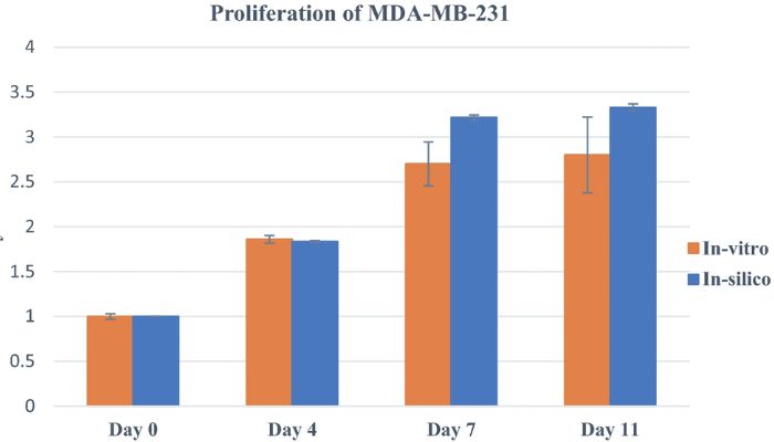
Graph to show the proliferation of the experimental cells in the mathematical model compared to the experimental results. The Y axis represents the number of proliferating cells. (Photo credit: Scientific Reports (Sci Rep))
In-silico refers to experiments which use or integrate computer models (in-silico being pseudo-Latin to mean the silicon chips in computers). For this research, the scientists used cellular automata modeling (modeling using a regular grid of cells), in which most of the parameters were picked from experimental data. A comprehensive in-vitro assay was also done to improve the reliability of the model; that is, the cells were examined to determine their composition to indicate and improve the quality of the model. The final results suggested that the 3D bioprinting optimization using modeling can be used to design experiments to achieve results without needing to conduct assays. The simulated data consistently replicated experimental results for viability and proliferation.
More from the desk
Six other notes from the rest of the bench.
 Printers#3DStartup: Apis Cor, Creators of the 3D Printed HouseIf you’ve been on the internet in the last year, you may have seen the viral clip of a 3D printed house built in just 24 hours. Apis Cor are a Russian company who built a whole, functional house in…
 Software3D Model Generator Tripo AI Raises 3 Billion Yuan in Series B and Series B+ FundingOn September 1, 2026, the generative 3D company Tripo AI announced it had raised approximately 3 billion yuan in its Series B and Series B+ financing rounds, equivalent to roughly $446 million USD. The funding will help the Tripo team…
 News3D Hubs Report: Key Market Findings and Trends for 20193D Hubs has released its traditional quarterly trends report. 3D Hubs is the world’s largest network of manufacturing services. Looking back on 2018, the company admits that the most significant change was the evolution of 3D printing from an almost…
 NewsHP and GE Additive are now among the biggest sellers of 3D printersWith the recent unveiling of a study on the largest vendors of industrial 3D printers in 2017, there is no doubt that HP and GE Additive, the additive manufacturing subsidiary of General Electric, have upset the market, entering the list…
 GuidesHow To Celebrate Easter With 3D PrintingThe sun is out, temperatures are warming up and Easter will take place this Sunday! Changing every year, Easter Sunday takes place following the first full moon after the vernal equinox (March 20th), though it is always on a Sunday…
 NewsPrintParts Partners with MakerGirl to Bridge STEM Gender GapEven with the advances made in recent years, women continue to be disproportionately unrepresented across STEM (science, technology, engineering, and math) industries, and in leadership positions in the technology sector. In 1970, women made up a mere 8% of STEM…