
Revolutionizing Healthcare: Real-Time 3D Printing Directly Onto Moving Organs with Motion Capture Technology
In a monumental leap forward for medical science and additive manufacturing, researchers at the University of Minnesota (UMN) have achieved a truly groundbreaking feat: successfully utilizing motion-capture technology to 3D print directly onto dynamic, expanding organs. This innovative application, which repurposes technology typically associated with the dazzling special effects of the film industry, promises to revolutionize a multitude of medical practices. What was once considered a far-fetched concept just a few years ago is now a tangible reality, offering immediate and profound implications, such as aiding in the diagnosis and continuous monitoring of the lungs of patients affected by diseases like COVID-19. This breakthrough signifies a major paradigm shift in how we approach personalized medicine and complex surgical interventions.
While additive manufacturing has demonstrated its versatility across an extensive array of materials, ranging from robust plastics and high-performance metals to intricate biomaterials, its impact on bioprinting applications targeting live biological surfaces has historically been slower to materialize. The primary hurdle lies in the inherent difficulty of 3D printing directly onto moving, soft tissues. The human body is a dynamic system; living organs are never truly static. They continuously expand, contract, and subtly shift, presenting an immense challenge for precise deposition. For a 3D printer to accurately deposit material, its sensors and deposition toolpath must adapt instantaneously to these constantly changing parameters, demanding an unprecedented level of real-time responsiveness and precision. Traditional 3D printing methods, designed for static or rigidly constrained environments, simply could not cope with this biological dynamism. This limitation severely constrained the potential of bioprinting for *in vivo* applications, pushing researchers to seek radically new approaches.
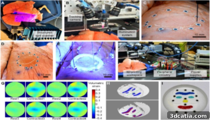
Image credits: Science Advances
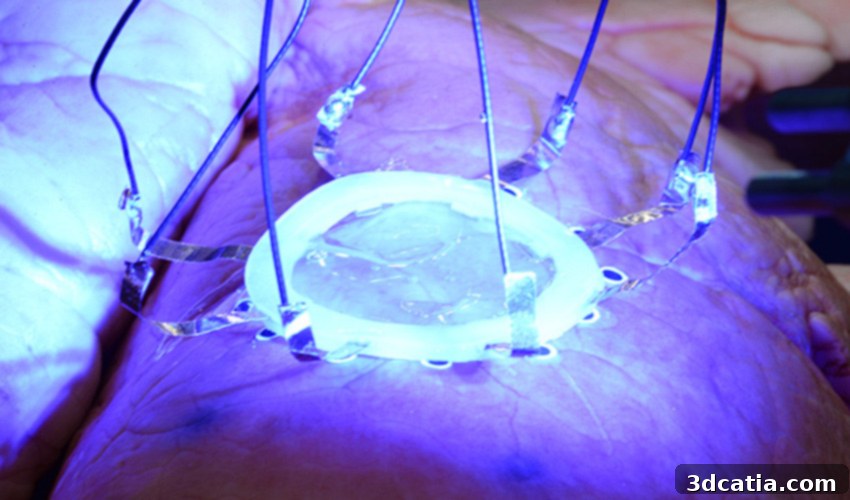
The UMN researchers meticulously addressed this formidable challenge by developing an ingenious new technique. Their methodology leverages a sophisticated dual-camera system, which works in conjunction with motion capture technology, to generate a dynamic toolpath for the 3D printer in real time. The core principle revolves around the idea that the complex “space of deformation” of a biological surface can be meticulously “learned” and predicted from a comprehensive dataset of 3D scans. By continuously tracking the organ’s movement, this system allows for the accurate recovery of its surface geometry in three dimensions. This real-time geometrical data is then fed directly back to the 3D printer, enabling it to instantaneously adapt its printing trajectory to the organ’s expanding and contracting motions. To achieve this, the team employed motion capture tracking markers – precisely the same type of markers that actors wear in Hollywood productions to create believable special effects. These markers provide the critical data points that allow the system to map and predict the organ’s movements with exceptional fidelity. To provide a compelling demonstration of their novel sensor and motion capturing technique, the research team successfully 3D printed a hydrogel-based Electrical Impedance Tomography (EIT) strain sensor directly onto a breathing lung, showcasing its unprecedented ability to monitor organ deformation with remarkable accuracy.
While the initial results are exceptionally promising, the researchers acknowledge that further refinements are necessary. Key areas for improvement include enhancing the biocompatibility of the printed sensors and boosting the overall precision of the technique. Once these advancements are achieved, this adaptive 3D printing method is poised to unlock a vast new frontier of surgical applications for bioprinting, fundamentally transforming how medical procedures are conducted. Consider clinical scenarios that currently require repeated, often imprecise, injections of biological materials. The advent of *in situ* autonomous 3D printing could entirely supersede manual operations, offering unparalleled spatial control over the deposition of materials for extended durations. This capability would not only enhance accuracy but also minimize invasiveness and reduce the risk of complications, paving the way for more effective and patient-specific treatments. This technology can enable the localized delivery of drugs, growth factors, or even stem cells directly to damaged or diseased tissues within the body, ensuring optimal therapeutic concentrations at the target site.
Looking ahead, it is highly probable that this adaptive 3D printing technique will play a pivotal role in augmenting robot-assisted medical treatments. By integrating real-time additive manufacturing capabilities, surgical robots could gain the ability to autonomously and precisely print biological materials directly onto or even inside the human body during complex procedures. This integration would not merely automate tasks but elevate surgical precision to an unprecedented level, allowing for customized tissue repair, personalized drug patches, or even the creation of intricate biological scaffolds *in vivo*. Professor Michael McAlpine, the distinguished lead researcher on this groundbreaking project, encapsulates this visionary future, stating, “We are pushing the boundaries of 3D printing in ways we never even imagined years ago. In the future, 3D printing will not be just about printing, but instead be part of a larger autonomous robotic system. This could be important for diseases like COVID-19, where health care providers are at risk when treating patients.” His words underscore the profound potential for this technology to safeguard medical professionals, streamline healthcare delivery, and fundamentally redefine patient care, especially in challenging environments or during public health crises.
The implications of this breakthrough extend far beyond immediate surgical applications. Imagine a future where personalized implants, tailored to the exact contours and biomechanics of a patient’s body, can be printed directly during an operation. Or where sensors are autonomously printed onto organs to provide continuous, real-time diagnostic information, allowing for early detection of disease progression or treatment response. This level of precise, on-demand fabrication opens up incredible possibilities for regenerative medicine, allowing for the repair or even replacement of damaged tissues with unprecedented accuracy. The ability to “print” functional components or therapeutic agents precisely where and when they are needed within a dynamic biological environment represents a monumental step towards truly personalized and adaptive healthcare. It promises to enhance patient outcomes, reduce recovery times, and introduce a new era of minimally invasive yet highly effective treatments. The integration of artificial intelligence and machine learning with this motion-capture 3D printing system could further refine its predictive capabilities, making the printing process even more robust and reliable in diverse clinical scenarios.
This pioneering work from the University of Minnesota marks a significant inflection point in the journey of medical technology. By bridging the gap between advanced manufacturing, robotics, and biology, these researchers are not just improving existing medical procedures; they are creating entirely new paradigms for diagnosis, treatment, and surgical intervention. The vision of a healthcare system where autonomous robotic platforms, equipped with real-time adaptive bioprinting capabilities, can perform intricate procedures with superhuman precision is rapidly moving from the realm of science fiction to a tangible reality. This will undoubtedly lead to safer, more effective, and more accessible healthcare solutions for a global population facing ever-evolving medical challenges. The precision offered by such a system could also minimize collateral damage to healthy tissues, a common concern in many surgical procedures.
What are your thoughts on this incredible motion capture technology being used for real-time 3D printing directly onto expanding organs? The potential for medical innovation is truly boundless. Share your insights and predictions in a comment below, or join the conversation on our vibrant Facebook and Twitter pages! Don’t miss out on the latest advancements and news in the world of additive manufacturing. Sign up for our free weekly Newsletter to get all the cutting-edge updates in 3D printing delivered straight to your inbox!