
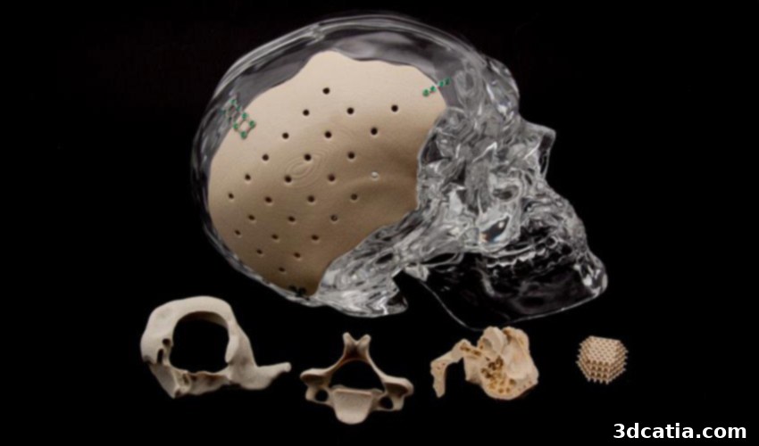
Revolutionizing Bone Regeneration: How 3D Printed PEKK Implants and Stem Cells Accelerate Healing
The medical field is witnessing a profound transformation driven by advancements in high-performance materials and additive manufacturing. Specifically, polymers such as PEEK, ULTEM, and PEKK are becoming indispensable for intricate medical applications, particularly due to their exceptional biocompatibility, strength, and sterilizability. These properties make them ideal for creating implants that integrate seamlessly with the human body. While earlier innovations have showcased the potential of these materials, for instance, the groundbreaking 3D printed clavicle made from PEEK in China, a new breakthrough promises to push the boundaries of regenerative medicine even further. Recent research from American institutions has now provided compelling evidence that 3D printed PEKK implants can significantly accelerate bone regeneration when combined with advanced stem cell therapies, offering a novel solution for patients with severe bone defects.
This pioneering study, a collaborative effort between researchers at Oxford Performance Materials (OPM) in Connecticut and McGill University in Canada, has been rigorously peer-reviewed and published in the prestigious scientific journal Nature. Their innovative approach focuses on enhancing the regenerative capacity of PEKK implants through biological augmentation. The researchers found that when 3D printed PEKK implants were combined with human synovial fluid mesenchymal stem cells (hSF-MSCs), the results were remarkable. Human synovial fluid, typically found in joint cavities, is a rich source of these potent mesenchymal stem cells, which possess a unique ability to regenerate cartilage and, crucially for this study, strongly promote new bone growth. This synergistic combination – a biocompatible, geometrically customized PEKK scaffold acting as a framework for cellular proliferation and differentiation – demonstrates immense potential. It could fundamentally change the treatment paradigm for individuals with severe bone defects, drastically reducing the reliance on more invasive and complex procedures such as traditional bone transplants, which often carry significant risks and limitations.
Bone transplants, whether autologous (from the patient’s own body) or allogeneic (from a donor), are standard procedures for replacing missing or damaged bone. An autologous bone transplant, for instance, involves surgically removing a section of healthy bone from one part of the body, such as the hip or fibula, and transplanting it to the affected area. This procedure, though effective in many cases, is fraught with potential complications. Worldwide, an astounding 2.2 million bone graft operations are performed annually, addressing a wide array of conditions from orthopedic trauma and reconstructive surgery to complex dental procedures. However, traditional bone transplantation carries considerable drawbacks, including prolonged regeneration times, potential infection at both the donor and recipient sites, and complications associated with the removal of autologous bone, such as chronic pain, nerve damage, or a weakening of the donor limb. The supply of donor bone can also be limited, and the potential for immune rejection exists with allogeneic grafts. Given these significant limitations and the immense patient burden, scientists and medical practitioners are actively seeking more efficient and safer alternatives. The study authors emphasize that bone regeneration using innovative biomaterials and stem cells represents the most promising approach to circumvent these long-standing challenges, offering a pathway to superior healing outcomes and reduced patient morbidity.
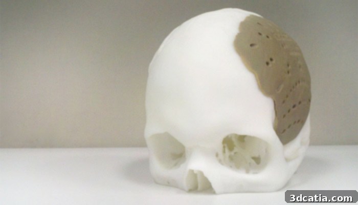
An advanced skull implant made of OXPEKK material, demonstrating the intricate possibilities of 3D printing in medical applications. | Credits: Oxford Performance Materials.
To rigorously evaluate the efficacy of their innovative strategy, the research team undertook a detailed investigation, exploring various cell types and optimizing biomimetic scaffolds specifically engineered for robust bone regeneration. Their methodology involved state-of-the-art additive manufacturing processes. The PEKK implants themselves were produced using the highly precise EOSINT P800 SLS printer, developed by the renowned German manufacturer EOS. This advanced Selective Laser Sintering (SLS) technology is crucial for creating complex geometries with controlled porosity, which is vital for enabling cellular ingrowth and nutrient diffusion within the implant structure. The effectiveness of these engineered PEKK implants, synergistically combined with hSF-MSCs, was then demonstrated *in vivo* through a compelling experiment: using the technology to heal a significant skull bone defect in a rabbit model. Animal models are indispensable in preclinical research, providing a vital bridge between laboratory discoveries and potential human applications, allowing for the observation of biological interactions and regenerative processes in a living system.
The outcomes of the study were exceptionally encouraging and validated the profound potential of this novel approach. A paramount finding was the outstanding biocompatibility exhibited by the PEKK implants. Critically, there was no evidence of rejection by the host body’s immune system, nor were there any signs of inflammation or adverse tissue reactions at the implant site. This excellent integration with the surrounding biological tissues is a fundamental requirement for any successful medical implant. Furthermore, the meticulously designed physical characteristics of the 3D printed PEKK implants played a pivotal role in their regenerative success. The implants were engineered with a deliberately rough surface topography and an intricate network of open micropores. These structural features are far more than superficial; they are bio-functional. The rough surface provides an optimal anchoring point, facilitating superior adhesion and proliferation of the hSF-MSCs. Concurrently, the interconnected micropores create a vascular-friendly environment, allowing for the deep penetration of cells, blood vessels, and essential growth factors into the implant. This architecture effectively mimics the natural extracellular matrix of bone, providing a highly conducive scaffold that significantly promotes rapid cell growth, differentiation, and ultimately, accelerated formation of new, healthy bone tissue directly within and around the implanted structure.
The unequivocal conclusion drawn by the researchers highlights that PEKK implants, particularly when bio-activated with hSF-MSCs, represent an extremely promising and potentially transformative strategy for addressing large bone defects. This combined approach offers a superior, less invasive, and more biologically active alternative to conventional bone grafting techniques, effectively circumventing many of their inherent limitations and risks. The specific high-performance PEKK material utilized in this groundbreaking study is known as OXPEKK®, a proprietary formulation developed and manufactured by Oxford Performance Materials (OPM). OPM leverages its advanced OsteoFab® technology to process this unique polymer into patient-specific implants. This sophisticated additive manufacturing platform enables the creation of highly customized, anatomically precise geometries that perfectly match the individual patient’s defect, optimizing both the fit and the regenerative potential. This confluence of advanced material science, precision 3D printing, and cutting-edge stem cell biology is setting a new standard in regenerative medicine, promising faster recoveries, reduced patient discomfort, fewer complications, and significantly improved long-term outcomes for countless individuals suffering from complex bone injuries worldwide.
The long-term implications of this research extend far beyond the immediate clinical applications for bone repair. This breakthrough signifies a paradigm shift towards personalized regenerative medicine, where 3D printed biocompatible materials serve not just as inert replacements but as active biological scaffolds that intelligently guide and accelerate the body’s intrinsic healing processes. The ability to regenerate substantial bone volume without the need for additional surgical procedures to harvest donor bone significantly reduces patient trauma, minimizes hospital stays, and potentially lowers overall healthcare costs, all while enhancing clinical effectiveness. As additive manufacturing technologies continue their rapid evolution and our understanding of cellular biology deepens, we can anticipate further innovations and broader applications of such personalized regenerative therapies across various medical disciplines. This study not only provides a concrete, advanced solution for significant bone defects but also lays a robust foundation for future endeavors in tissue engineering, potentially impacting the repair and regeneration of other critical tissues and organs throughout the human body. The journey from pioneering research to widespread clinical adoption will undoubtedly involve extensive further testing, rigorous regulatory approvals, and sustained collaborative efforts among material scientists, biomedical engineers, and medical professionals, but the transformative potential demonstrated by OPM and McGill University is undeniably profound.
What are your thoughts on this revolutionary research that combines 3D printed medical implants with advanced stem cell therapies for accelerated bone regeneration? We would love to hear your perspectives and engage in a discussion! Feel free to share your comments below, or join the conversation on our Facebook and Twitter pages. To stay completely up-to-date with all the latest advancements and news in the dynamic world of 3D printing, remember to sign up for our free weekly Newsletter, delivered straight to your inbox!
*Cover Photo Credits: Oxford Performance Materials