
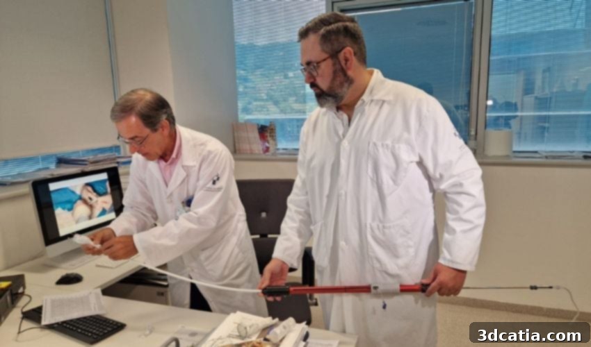
Revolutionizing Aneurysm Repair: HUCA Pioneers Custom 3D Printed Prostheses for Complex Abdominal Aortic Aneurysms
The Spanish Society of Cardiovascular and Endovascular Surgery highlights a critical concern: a substantial segment of the population over 60 faces a heightened risk of developing an abdominal aortic aneurysm (AAA). These potentially life-threatening lesions present a significant challenge for medical professionals, especially when they involve areas with branches leading to vital organs such as the kidneys, liver, and intestines. Traditionally, treating such complex cases has been akin to solving an intricate puzzle for surgeons. However, a groundbreaking solution has emerged from Oviedo, Spain, where the Central University Hospital of Asturias (HUCA) is leveraging the power of 3D printing technology to craft customized prostheses, enabling their implantation through innovative, minimally invasive procedures. This pioneering approach is not only transforming vascular surgery but also offering a new beacon of hope for patients with complex anatomical challenges.
Understanding Abdominal Aortic Aneurysms and Their Treatment Challenges
At its core, an aneurysm is an abnormal bulge or ballooning that forms in the wall of an artery. In the case of an abdominal aortic aneurysm, this enlargement occurs in the aorta, the body’s largest artery, which originates from the heart and descends through the chest and abdomen, supplying blood to the rest of the body. An AAA is particularly dangerous because if it ruptures, it can lead to massive internal bleeding, which is often fatal. The risk of developing an AAA increases significantly with age, and factors such as smoking, high blood pressure, and a family history of aneurysms also contribute to its prevalence.
The challenge of treating an AAA intensifies dramatically when the aneurysm is juxtarenal or pararenal – meaning it is located very close to or involves the arteries that supply blood to critical visceral organs like the kidneys, liver, and intestines. In such intricate anatomies, any intervention must meticulously maintain blood flow to these vital organs while simultaneously reinforcing the weakened, damaged aortic artery. This dual requirement makes these cases exceptionally complex and risky.
Historically, open surgery has been the primary method for repairing AAAs. This involves a large abdominal incision, clamping the aorta, and replacing the affected section with a synthetic graft. While effective, open surgery is a highly invasive procedure associated with significant risks, including substantial blood loss, infection, lengthy hospital stays, and extended recovery periods. For elderly patients or those with existing comorbidities such as heart disease, lung conditions, or kidney problems, the risks associated with open surgery can be prohibitively high.
The advent of endovascular aneurysm repair (EVAR) offered a less invasive alternative. EVAR involves inserting a stent-graft through small incisions (typically in the groin) and navigating it to the site of the aneurysm. The stent-graft then expands to reinforce the arterial wall, preventing rupture. However, standard, off-the-shelf endovascular prostheses are designed for more common anatomical configurations. In complex cases where the aneurysm involves critical branch arteries, these standard grafts often fall short. They may not provide an adequate seal, leading to endoleaks (blood flowing outside the graft), or they might block blood flow to essential organs, necessitating further, often complex, interventions. Achieving a precise, secure fit in these highly irregular and tortuous anatomies has been a persistent limitation of conventional EVAR, leaving many patients with complex AAAs facing the high risks of open surgery or no viable treatment option.
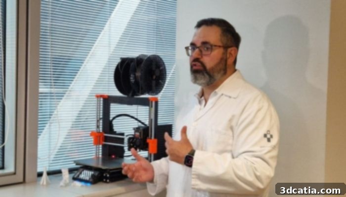
Dr. Francisco Álvarez Marcos, Associate in the Angiology and Vascular Surgery Department at the Central University Hospital of Asturias (HUCA)
The HUCA Innovation: Leveraging 3D Printing for Patient-Specific Solutions
To overcome these significant limitations and enhance patient outcomes, a visionary team from the Angiology and Vascular Surgery Department at HUCA, spearheaded by Dr. Manuel Alonso, turned to cutting-edge 3D printing technology. Their innovative methodology centers on creating truly customized endovascular prostheses, precisely tailored to each patient’s unique vascular anatomy. This patient-specific approach marks a paradigm shift in the treatment of complex abdominal aortic aneurysms.
The process begins with highly detailed imaging – typically a computed tomography (CT) angiography scan – which captures the intricate geometry of the patient’s aorta and its branching vessels. This high-resolution data is then used to generate an accurate digital 3D model of the patient’s anatomy. Subsequently, this digital model is brought to life as a physical, tangible replica using a 3D printer. This 3D printed anatomical model serves as an invaluable tool for surgical planning, allowing the medical team to visualize and interact with the patient’s exact vascular structure in a way that 2D images cannot provide. It precisely reproduces the “normal” blood flow path, enabling meticulous pre-operative assessment and strategy formulation.
The true innovation lies in the subsequent step: on this physical 3D printed model, the surgical team modifies a conventional, off-the-shelf endovascular prosthesis. Using the model as a guide, they meticulously create custom fenestrations (openings) and scallops (indentations) in the graft, aligning them perfectly with the exact locations where the visceral arteries (renal, superior mesenteric, celiac) branch off the aorta. This ensures that once implanted, the prosthesis will reinforce the aneurysm without compromising blood flow to these critical organs. This level of precision customization, previously unattainable, is a game-changer.
Before entering the operating room, an additional critical verification step is performed: the customized design is checked using a paper template that mimics the exact structure of the modified prosthesis. This ‘dress rehearsal’ significantly reduces the potential for errors during the actual surgery and ensures a flawless, precise fit. The paramount advantage of this entire process is achieving a level of customization comparable to or even surpassing that of expensive, conventionally custom-manufactured prostheses, but in a significantly reduced timeframe and at a lower cost, all while being perfectly tailored to the individual patient’s anatomy. This bespoke approach minimizes the risk of complications associated with imperfect graft sizing or positioning, such as endoleaks or renal ischemia, which have historically plagued complex EVAR procedures.
Minimally Invasive Procedure and Remarkable Outcomes
The implementation of these custom 3D printed prostheses allows for a highly effective minimally invasive endovascular procedure. Unlike open surgery, which necessitates large incisions, this innovative technique is performed with only small skin punctures, typically in the groin where catheters are inserted into the femoral arteries. The custom-designed graft is then carefully guided through the arterial network to the aneurysm site and deployed. This approach drastically reduces surgical trauma to the patient.
The benefits for patients are profound. The procedure usually lasts less than three hours, a considerable reduction compared to complex open surgeries. The minimally invasive nature facilitates a significantly faster recovery period, allowing patients to be discharged from the hospital much sooner and resume their daily activities more quickly. Furthermore, it substantially reduces the risk of post-operative complications, including infection, pain, and blood loss, which are common concerns in traditional open surgery, especially for older patients or those with multiple health issues.
The initial results from HUCA’s pioneering work are exceptionally promising. The first 18 patients treated with this method had an average age of 80, highlighting the suitability and safety of the technique for a vulnerable, elderly population. In total, 37 critical blood vessels were successfully treated, demonstrating the technique’s efficacy in managing complex branch artery involvement. The technical success rate achieved was an impressive 94%, meaning the prostheses were accurately positioned and functioned as intended in almost all cases. Crucially, there was no mortality reported in the first month following the procedure, a remarkable achievement for such high-risk interventions. Furthermore, none of the patients required additional interventions within this period, and a 100% patency rate was observed in the repaired arteries, confirming sustained blood flow to the vital organs. These outcomes underscore the safety, precision, and enduring effectiveness of HUCA’s 3D printing-enabled approach.
The groundbreaking technique and its compelling results have garnered international recognition. They were presented at the prestigious VEITH Symposium in New York, a leading global forum for vascular specialists, and subsequently published in the esteemed journal, Journal of Endovascular Therapy, solidifying its place in the medical literature and affirming its scientific validity.
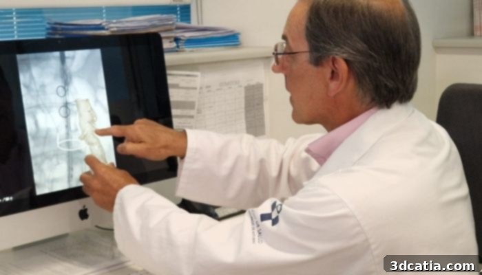
Dr. Manuel Alonso from the Angiology and Vascular Surgery Department at the Central University Hospital of Asturias (HUCA)
Pioneering the Future of Vascular Surgery
HUCA’s innovative approach stands as a powerful testament to how advanced 3D technologies are fundamentally transforming medical fields, particularly vascular surgery, where we are witnessing an accelerating adoption of these applications. The ability to create highly detailed, patient-specific anatomical models and custom implants represents a significant leap forward in personalized medicine. This not only enhances surgical planning and execution but also paves the way for a future where every patient receives treatment meticulously tailored to their unique physiological needs.
With these very positive initial results, the 3D printing-assisted technique looks exceptionally promising, especially given its distinct advantages in safety, precision, and speed compared to conventional methods. It offers a viable and superior treatment option for patients previously considered high-risk or untreatable with standard endovascular grafts, thereby expanding the reach of minimally invasive procedures. This innovative work at HUCA exemplifies the potential of additive manufacturing to improve patient care, reduce healthcare burdens, and ultimately save lives by addressing some of the most complex challenges in modern medicine.
To delve deeper into the specifics of this groundbreaking technique, interested readers can consult the official press release from the Principality of Asturias HERE.
Join the Conversation on Medical 3D Printing
What are your thoughts on the revolutionary use of 3D printing at HUCA for treating complex abdominal aortic aneurysms? We invite you to share your insights and comments below, or engage with us on our LinkedIn or Facebook pages! Don’t miss out on the latest advancements in additive manufacturing – sign up for our free weekly Newsletter to receive top 3D printing news directly in your inbox. You can also explore our extensive video content on our YouTube channel. For more news and developments specifically in medical and dental 3D printing, be sure to visit our dedicated page HERE.
*All Photo Credit: Principado de Asturias