

Revolutionizing Neurological Repair: Oxford University Pioneers 3D Bioprinted Brain Tissue for Traumatic Brain Injury
Traumatic brain injury (TBI) represents a formidable global health challenge, impacting an estimated 70 million individuals worldwide annually. These injuries, often resulting from head trauma, range from mild concussions to severe conditions that can lead to permanent disability or even be fatal. For decades, the scope of effective treatments for TBI has remained profoundly limited, leaving many patients with lifelong impairments and little hope for significant recovery. However, a groundbreaking new study from Oxford University researchers signals a potential paradigm shift in neurological repair. Their recently published research details the successful 3D bioprinting of viable brain tissue, which demonstrated remarkable integration with existing host brain tissue in rigorous laboratory tests. This pioneering achievement underscores the immense potential that 3D bioprinting could bring to the treatment of brain injuries, offering a beacon of hope where conventional medicine has largely stalled.
This monumental development from Oxford builds upon a burgeoning field of regenerative medicine and follows closely on the heels of other significant advancements, such as Monash University’s breakthrough in 3D printed nerve cells. The Oxford team’s extensive background in bioprinting research has culminated in this innovative approach, which introduces a new dimension to the study of 3D printing for tissue replacements. By skillfully utilizing neural stem cells, the bioprinted tissue was engineered to meticulously replicate the intricate biological architecture of a cerebral cortex, the brain’s outermost layer responsible for higher cognitive functions. A persistent challenge for researchers in bioprinting has been the consistent and effective mimicking of desired tissue or organ structures. This study, however, vividly illustrates that through the strategic incorporation of guiding components, such as patient-derived neural stem cells, 3D printed brain tissue can exhibit greater cooperation and facilitate easier integration within the body. The ability to derive these stem cells directly from the patient themselves offers a crucial advantage, significantly reducing the risk of immune rejection and paving the way for personalized regenerative therapies.
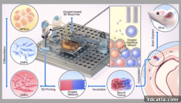
Diagram of the 3D bioprinter and the materials that make up the bioink (Image credits: Jin et al. / University of Oxford)
The methodology employed in this pioneering research involves a technique known as ‘droplet printing.’ This process shares fundamental similarities with other advanced additive manufacturing techniques that rely on an inkjet printhead, such as material jetting. In droplet printing, precise micro-droplets of bioink are deposited layer by layer to construct the desired biological structure. For this specific study, the researchers carefully combined various essential materials, including human induced pluripotent stem cells (hiPSCs), into two distinct bioinks. These specialized bioinks were then utilized to meticulously create dual layers that remarkably resembled the complex structure of a cerebral cortex. A critical success factor highlighted by the research was the printed tissue’s ability to maintain its structural integrity and form for an extended period, even when subjected to storage conditions. This stability is paramount for potential clinical applications, ensuring that the bioprinted tissue can be handled, transported, and stored effectively before implantation. The precision and control offered by droplet printing, combined with the carefully formulated bioinks, were instrumental in achieving the anatomical fidelity and long-term viability observed in the synthetic brain tissue.
Following the successful conclusion of this initial phase of the study, the senior authors of the research paper, representing diverse departments including Chemistry and Physiology, Anatomy, and Genetics, shared their insights and perspectives. Their collective sentiment underscored both the profound complexity of human brain development and the significant strides made by their team. Professor Zoltán Molnár eloquently articulated this balance, stating, “Human brain development is a delicate and elaborate process with a complex choreography. It would be naïve to think that we can recreate the entire cellular progression in the laboratory. Nonetheless, our 3D printing project demonstrates substantial progress in controlling the fates and arrangements of human iPSCs to form the basic functional units of the cerebral cortex.” This statement reflects a pragmatic yet highly optimistic view, acknowledging the immense intricacy of the brain while celebrating the crucial steps taken toward building its fundamental components. The interdisciplinary nature of the research, bringing together expertise from various scientific fields, was undoubtedly a key factor in achieving such sophisticated control over stem cell differentiation and arrangement, enabling the creation of structures that closely mimic the brain’s natural architecture.
Buoyed by their initial success, the Oxford team is now poised to embark on the next ambitious phase of their research. Their immediate plans include further refining the intricate printing process, aiming to introduce additional layers and increased biological complexity into the bioprinted tissues. The ultimate goal is to more accurately reflect the multifaceted structural and functional nuances of the human brain, bringing them closer to creating fully functional neural implants. Beyond the direct treatment of brain injuries, the potential applications for this pioneering research extend into several vital avenues of neuroscience and medicine. These include revolutionary drug testing platforms, where patient-specific brain tissues could be used to evaluate new pharmaceuticals with unprecedented accuracy, reducing reliance on animal testing and accelerating drug discovery for neurological conditions. Additionally, the bioprinted brain models could serve as invaluable tools for studying brain development, offering insights into how neurological disorders arise and progress, and potentially identifying early intervention strategies. Furthermore, this technology opens new frontiers for fundamental research into our understanding of cognition itself, allowing scientists to model and investigate complex neural networks in a controlled laboratory setting. This multi-faceted approach signifies a future where 3D bioprinting not only repairs damaged brains but also vastly expands our knowledge of the most complex organ in the human body. For those interested in delving deeper into the specifics of this groundbreaking research, the full paper is available HERE.
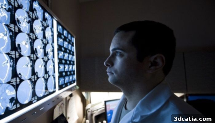
Bioprinting may prove to be a revolutionary way to treat brain injuries (Photo credits: USAF)
The advancements made by Oxford University in 3D bioprinting brain tissue herald a new era in neurological medicine, offering unprecedented hope for those affected by traumatic brain injuries and a multitude of other neurological conditions. The ability to create functional, integrative brain tissue promises to transform treatment paradigms, pushing the boundaries of what is medically possible. We are eager to hear your thoughts on the profound potential medical uses of 3D printed brain tissue. Let us know your perspective in a comment below or engage with us on our LinkedIn, Facebook, and Twitter pages! Don’t forget to sign up for our free weekly Newsletter here to receive the latest 3D printing news straight to your inbox! You can also find all our compelling videos and interviews on our YouTube channel, where we explore the cutting edge of additive manufacturing.
*Cover Photo Credits: University of Oxford