

Revolutionizing COVID-19 Understanding: How 3D Printing Creates Life-Size Lung Models for Enhanced Patient Care and Medical Insight
As the world continues to grapple with the profound and multifaceted challenges posed by the COVID-19 global pandemic, medical professionals and researchers are relentlessly working to unravel the complexities of this novel disease. A critical concern that has emerged from the initial waves of infection is the potential for severe, long-term complications, particularly affecting the respiratory system. Doctors have issued warnings regarding the necessity of prolonged treatment for COVID-19 survivors, primarily due to the virus’s propensity to induce severe pneumonia, leading to significant and often critical damage to the lungs. Therefore, equipping medical teams with an unparalleled understanding of the virus’s physiological impact is paramount to developing and delivering optimal treatment pathways for patients. In this endeavor, innovative medical technology companies are playing a crucial role, with Axial3D, a pioneer in 3D printing technologies for healthcare, stepping up to provide invaluable insights.
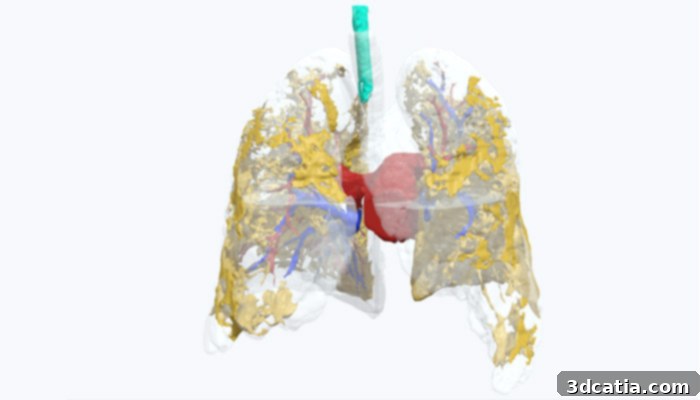
Axial3D embarked on a significant collaborative project with the dedicated respiratory team at the Belfast Health and Social Care Trust. This partnership aimed to create a highly precise, 1:1 scale 3D anatomical model of a real COVID-19 patient’s lungs. The process began with advanced CT scans, which were captured on the 14th day following the patient’s infection. These detailed scans provided the foundational data necessary to accurately reproduce the complex internal structures of the lungs. Once the digital 3D model was meticulously prepared from the radiological data, Axial3D leveraged Formlabs’ cutting-edge Stereolithography (SLA) technology. This highly accurate additive manufacturing process allowed them to produce the physical scale model on a Form 3 3D printer. To facilitate an unprecedented level of detailed examination, the lungs were printed in eight separate sections. These individual parts were then ingeniously joined together using precision-placed magnets, allowing for easy assembly and disassembly. This modular design proved instrumental, as it enabled medical experts to explore a comprehensive range of views, visualizing the precise depth and distribution of the infection within the lung tissue. Such a detailed, multi-dimensional perspective delivers unique and critical insights into how the virus infiltrates and impacts the patient’s respiratory system, moving beyond the limitations of traditional 2D imaging.
The utility of 3D printing technologies in the medical sector is expansive and transformative, spanning a vast array of applications that fundamentally enhance patient care and medical understanding. From the creation of highly customized medical devices, such as bespoke prosthetics and orthotics tailored to individual patient anatomy, to the production of intricate surgical guides that improve precision in complex procedures, the technology empowers healthcare professionals. Crucially, it enables the rapid and cost-effective creation of precise, patient-specific anatomical models, like the COVID-19 lung model. This capability is precisely why Axial3D has strategically integrated 3D printing into its core operations, specializing in making this advanced technology widely accessible to the entire healthcare sector. For patients battling COVID-19, and for the clinicians striving to understand and mitigate its effects, this level of specialized expertise in precision medical modeling is an invaluable asset, offering a tangible means to visualize, plan, and educate in an unprecedented way.
Dr. Paul McKeagney, a frontline physician who has treated numerous COVID-19 patients, including some of the most severely affected individuals in Northern Ireland, offered profound insights gleaned from the 3D printed lung model. He specifically drew attention to certain yellow areas visible within the model. Dr. McKeagney meticulously explains the clinical significance of these colored sections: “The yellow material inside the model represents pus, which is the body’s natural inflammatory response to the infection. This accumulated pus occupies regions of the lungs that are typically hollow and optimally designed for vital gas exchange – the process where oxygen enters the bloodstream and carbon dioxide is expelled. When these critical air sacs become inundated with pus and other watery inflammatory substances, the lungs’ fundamental ability to function properly is severely compromised. This impairment leads to profound respiratory distress and necessitates significant medical intervention and ventilatory support to aid the patient’s breathing.” The ability to physically see this internal pathology in a life-size, tangible format makes the abstract concept of lung damage profoundly clearer for clinicians and trainees alike, enhancing diagnostic understanding and treatment planning.
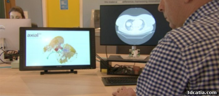
Image via BBC
One of the most compelling advantages of this real-life 3D anatomical model is its capacity to convey complex medical information with unparalleled clarity, even to individuals without specialized expertise in radiology. Traditionally, interpreting intricate CT scans requires years of dedicated training and experience. However, this tangible model democratizes understanding, allowing a broader range of medical professionals, students, and even patients and their families to grasp the extent and nature of the damage. Roger Johnston, CEO at Axial3D, eloquently captured the transformative impact of this innovation: “When discussing the severe lung pathology associated with COVID-19 with a radiologist, we proposed the innovative idea of 3D printing the affected lungs. The radiologist promptly provided us with comprehensive sets of CT scans, which we then meticulously processed and printed in our state-of-the-art Formlabs 3D print lab. The result has been nothing short of incredible; we are now able to visualize the internal structure and damage of the lungs in a manner that has truly never been seen before. This advancement has empowered the medical team with a completely new and profound way of viewing and understanding the complex, long-term effects of the infection.” This tangible visualization bridges the gap between abstract 2D images and the complex reality of human anatomy, fostering deeper comprehension and facilitating more informed clinical decisions.
The creation of such patient-specific 3D printed anatomical models offers a multitude of benefits across the healthcare spectrum. For multidisciplinary teams involved in patient care, these models serve as invaluable communication tools, enabling clearer discussions during ward rounds or pre-operative briefings, even for non-surgical cases like severe respiratory illness. They allow pulmonologists, intensivists, radiologists, and respiratory therapists to converge on a shared, tangible understanding of a patient’s condition, fostering collaborative decision-making. Furthermore, these models are exceptionally effective for patient and family education. Explaining complex medical diagnoses, particularly severe lung damage from a virus like COVID-19, can be challenging. A physical model provides a clear visual aid, helping patients and their families comprehend the extent of the disease, the rationale behind treatment plans, and the potential for long-term recovery. This enhanced understanding can significantly improve patient adherence to treatment and overall psychological well-being. Beyond direct patient care, these models are revolutionary for medical training and education, offering students and residents a hands-on learning experience that far surpasses what can be achieved with textbooks or even digital simulations alone, preparing them for real-world clinical scenarios.
Looking ahead, the successful application of 3D printing in understanding COVID-19 lung damage highlights its immense potential for addressing a broader range of medical challenges. This technology can be leveraged to create patient-specific models for other complex respiratory diseases, congenital heart defects, cancerous tumors, or intricate bone structures, offering unparalleled insights for diagnosis, surgical planning, and personalized treatment strategies. As healthcare systems globally face increasing demands and the potential for future pandemics, the ability to rapidly produce accurate anatomical models from medical imaging can accelerate research, improve diagnostic accuracy, and enhance preparedness for critical health crises. It represents a significant step forward in precision medicine, where treatment is tailored not just to the disease, but to the unique anatomical and physiological characteristics of each individual patient. This innovative use of 3D printing truly underscores how technological advancements can profoundly impact human health, fostering a new era of medical understanding and patient-centric care.
What are your thoughts on utilizing patient-specific, life-size 3D printed lung models to deeply study the complex effects of COVID-19 and advance medical understanding? We encourage you to share your perspectives in a comment below or join the conversation on our Facebook and Twitter pages! For all the latest news and innovative applications in 3D printing, sign up for our free weekly Newsletter, delivered straight to your inbox.