
Unlocking Ancient Worlds: 3D Scanning and Printing Revolutionize Museum Collections and Research
Museums worldwide are undergoing a profound transformation, actively embracing cutting-edge technologies to redefine and enhance access to their invaluable artifact collections. In this exciting era of digital innovation, two technologies stand out as particularly transformative: 3D scanning and 3D printing. These powerful tools are not merely modern conveniences; they represent a paradigm shift, offering unprecedented opportunities for researchers, educators, and enthusiasts alike to engage with historical, scientific, and cultural specimens in truly immersive and interactive ways. From fragile fossils to intricate ancient tools, the ability to create precise digital replicas is opening up new frontiers in preservation, study, and public engagement, ensuring that our shared heritage can be explored without compromising the original artifacts.
At the vanguard of this technological revolution is the University of California, Berkeley’s Museum of Vertebrate Zoology (MVZ). The MVZ is home to an astonishing collection of over 300,000 meticulously preserved reptile and amphibian vertebrate specimens, each holding a unique story about our planet’s biodiversity and evolutionary history. Recognizing the immense potential of digital technologies, Berkeley has spearheaded the openVertebrate project (oVert) in collaboration with more than 25 other prestigious museums across the United States. This ambitious initiative is fundamentally driven by a critical dual mission: to digitize these vast and often fragile collections through advanced 3D scanning techniques, thereby ensuring their long-term preservation while simultaneously dramatically expanding their accessibility to a global audience of researchers, students, and the general public. The inherent challenges of preserving delicate biological specimens while making them available for rigorous scientific study are immense, and oVert offers a groundbreaking, scalable solution.
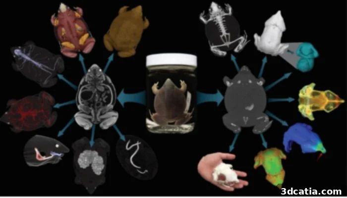
MVZ 3D scans of amphibian vertebrate, muscle tissue, skeleton, and more achieved through CT scans and X-rays.
The seamless integration of high-resolution 3D scanning has been a pivotal factor in the remarkable success and rapid expansion of the oVert project. By enabling the swift and extensive digitization of vertebrate collections nationwide, this technology has created an unparalleled digital archive that is both comprehensive and easily navigable. Scientists are now leveraging these incredibly detailed scans, often obtained through advanced CT scans and X-rays, to conduct intricate anatomical studies with a precision previously unimaginable. These digital models provide a wealth of crucial evolutionary and ecological data, allowing researchers to explore minute anatomical features, internal structures, and comparative morphologies without ever physically handling the delicate original specimens. This capability is particularly vital in zoological research, where museums have a profound responsibility to carefully consider the ethical implications of collecting animals from the wild. The specimens gathered must provide significant value for both current and future scientific inquiry, and 3D digitization maximizes this value.
With 3D scanning, a transformative new avenue has emerged for studying and accessing these irreplaceable specimens. It offers the profound advantage of zero risk of degradation, damage, or wear and tear to the original physical artifacts, a perennial concern with repeated physical handling, especially for fragile biological materials. Researchers can virtually manipulate, dissect, and analyze digital models from anywhere in the world, fostering global collaboration and accelerating scientific discovery without the need for costly and risky specimen transport. Furthermore, this digital approach significantly reduces the logistical and financial burdens associated with transporting physical specimens for study, making high-value research more accessible to a broader scientific community, including institutions with limited resources. The ability to zoom, rotate, and even ‘slice’ through a virtual model offers insights that might be impossible or too damaging to achieve with a physical specimen, truly pushing the boundaries of anatomical, paleontological, and ecological research.
Berkeley’s Vision for Enhanced Museum Access and Global Engagement
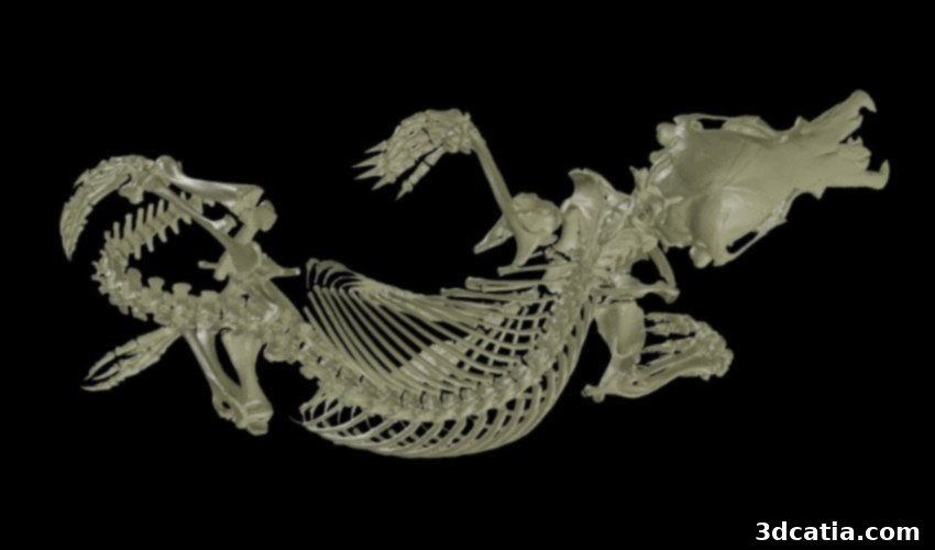
A compelling and highly illustrative example of the transformative impact 3D scanning is having within museums is the remarkable digital resurrection of a juvenile Australian platypus, *Ornithorhynchus anatinus*. This particular specimen, a rare and fascinating monotreme, has been carefully preserved in ethanol and stored at the MVZ for decades. While the physical artifact is, by its very nature and fragility, very rarely put on public display or loaned out for study, its meticulously digitized 3D scan has attracted an astonishing level of interest since its online release. The platypus model has quickly ascended to become the second most downloaded file within the entire oVert database, a powerful testament to the democratizing potential of digital accessibility and the public’s thirst for engaging with unique specimens.
Carol Spencer, the esteemed staff curator of herpetology in the MVZ, eloquently highlighted the profound shift in access and attention this artifact has gained. She shared a powerful anecdote that vividly illustrates this transformation: “We’ve had this platypus in ethanol in a big tank, but it’s never been loaned out. The only people who have ever gone to look at this are people that come here to our collection; it’s maybe been looked at twice in its entire history here at MVZ. But in six years, it’s been downloaded 320 times.” This striking contrast underscores the democratizing effect of 3D digitization. What was once an almost invisible specimen, known to only a handful of specialists and locked away for physical preservation, has now become a globally accessible research and educational resource, viewed and studied hundreds of times over by a diverse audience, all without ever leaving its protective environment. This also significantly broadens the reach of the museum’s collection, aligning with its mission to serve the public good.
The benefits of digital artifact collections extend far beyond unprecedented access for scientific researchers to detailed anatomical and genetic data. These high-fidelity 3D models also catalyze new avenues for rich interdisciplinary collaboration and innovative educational practices. Artists, for instance, can leverage these incredibly accurate 3D models to craft incredibly lifelike replicas, breathing new artistic interpretations into scientific specimens and creating engaging exhibits. Crucially, schools and universities worldwide have enthusiastically embraced these digital assets, utilizing them for truly immersive and engaging teaching experiences in classrooms. Imagine students exploring the intricate skeletal structure of an amphibian or the internal organs of a reptile directly on their screens, manipulating the models, and fostering a deeper understanding that any textbook or static image could provide. Once these invaluable collections are meticulously scanned, their digital files become readily available. They can be swiftly downloaded online from specialized repositories like Morphosource or popular platforms such as Sketchfab, and then sent to a 3D printer for convenient and cost-effective physical access. This capability means that a classroom in a remote location can print a detailed replica of a rare specimen, transforming theoretical learning into tangible, hands-on discovery and significantly reducing barriers to educational resources.
Looking ahead, the MVZ has ambitious and visionary plans to further expand its digital footprint, aiming to digitize roughly 800,000 more artifacts within the museum’s extensive collections. This monumental undertaking will significantly amplify the public’s accessibility to a vast array of biological specimens, democratizing knowledge and fostering a deeper appreciation for biodiversity and natural history. Carol Spencer reflected on these ongoing efforts and offered profound insight into the future trajectory of the oVert project. She articulated the inherent tension often faced in museum work, stating, “You want to protect specimens, but you also want to have people use them – oVert is a way of reducing the wear and tear on samples while also increasing access, and it’s the next logical step in the mission of museum collections.” Her words underscore the delicate yet critical balance between rigorous conservation and widespread dissemination, a challenge that 3D scanning masterfully addresses. By providing virtual access and the option for physical replicas, museums can safeguard their irreplaceable originals for eternity while simultaneously fulfilling their educational and research mandates on an unprecedented global scale. This approach not only preserves our collective natural heritage but actively transforms it into an open, living resource for future generations of scientists, students, and curious minds, ensuring its relevance and impact endure. To delve deeper into the fascinating details of this pioneering project, including its methodologies and ongoing discoveries, click here.
The Future of Digital Cultural Heritage and Scientific Discovery
The impact of groundbreaking projects like oVert extends far beyond the immediate benefits of individual specimen digitization. They lay the groundwork for a future where museum collections are not confined to physical buildings but exist as dynamic, accessible digital archives, perpetually available to anyone with an internet connection. This vision promises to revolutionize fields from taxonomy and evolutionary biology to art and educational technology, fostering innovation across disciplines. Imagine a world where researchers in developing countries have immediate access to rare specimens housed in distant museums, enabling breakthroughs regardless of geographical or financial constraints. Or consider K-12 students who can interact with digital models of ancient life forms as an integral part of their science curriculum, fostering a deeper, more engaging understanding of natural history than ever before. 3D printing further amplifies this, allowing for tactile engagement with complex anatomical structures that might otherwise remain abstract concepts, bridging the gap between digital data and physical understanding.
Moreover, these ongoing digitization efforts contribute significantly to global conservation initiatives. By providing detailed, reproducible data on species, researchers can better understand biodiversity trends, track changes over extended periods, and inform more effective conservation strategies for endangered species and threatened ecosystems. The digital nature of these models also means they are impervious to environmental degradation, natural disasters, or geopolitical instability, offering an invaluable and resilient layer of security for our shared natural heritage. The collaborative framework established by oVert, bringing together numerous institutions across the United States, also serves as an exemplary model for future large-scale digitization projects across various disciplines, from archaeology to anthropology, ensuring that cultural and scientific treasures are not only preserved but actively made available and utilized by all for generations to come. This represents a true democratization of knowledge and an exciting new chapter for museums worldwide.
What are your thoughts on UC Berkeley’s groundbreaking use of 3D scanning in the oVert project? We invite you to share your insights and engage with us! Let us know in a comment below or connect with us on our LinkedIn, Facebook, and Twitter pages! Don’t miss out on the latest advancements in 3D printing and digital heritage – sign up for our free weekly newsletter here for the freshest news delivered straight to your inbox! You can also find all our compelling videos and documentaries on our dedicated YouTube channel.
*All Photo Credits: The Museum of Vertebrate Zoology (MVZ)