
Revolutionizing Gastric Ulcer Treatment: In Situ Bioprinting Directly onto the Stomach Wall
The landscape of bioprinting technologies has witnessed remarkable advancements in recent years, pushing the boundaries of what’s possible in the medical field. This innovative method involves the precise combination and layer-by-layer deposition of living cells and biomaterials. The primary goal is to engineer biomedical models that faithfully replicate the complex properties of natural, living tissues. Beyond creating models for research, a significant focus within bioprinting research is dedicated to the ambitious endeavor of fabricating functional human organs in three dimensions, aiming to address the critical shortage of donor organs. Today, we delve into a groundbreaking new application of 3D printing in medicine, one that holds immense promise for transforming the treatment of digestive system ulcers. This pioneering technique, recently developed by researchers, involves the direct bioprinting of regenerative materials onto the stomach wall from within the body. But how exactly does this revolutionary process work?
A team of visionary researchers at Tsinghua University in China has spearheaded the development of a novel prototype leveraging additive manufacturing of biomaterials to facilitate the healing of gastric wounds from an internal perspective. Their extensive findings were meticulously documented in a research paper titled “Preliminary engineering for in situ in vivo bioprinting: a novel micro bioprinting platform for in situ in vivo bioprinting at a gastric wound site,” which was subsequently published in the esteemed scientific journal, IOPscience. This publication marks a significant milestone, introducing a new paradigm in therapeutic approaches for internal injuries.
Direct Bioprinting for Gastric Ulcers: An In-Depth Look
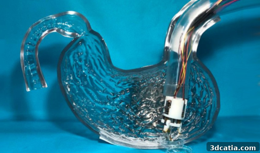
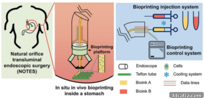
The core of this innovative treatment lies in a specially designed micro-robot, an engineering marvel crafted for precision and efficiency within the human digestive system. When folded for insertion, this compact device measures a mere 30mm in width and 43mm in length. However, once it reaches its designated target site and deploys, it expands to an impressive 59mm, providing a stable platform for its bioprinting operations. David Hoelzle, a distinguished mechanical engineer at Ohio State University, lauded the ingenuity behind this design, stating, “The team has designed ingenious mechanisms that enable a compact system to penetrate the body and, once the narrow entry restrictions are overcome, it deploys to cover a large work area.” This highlights the sophisticated mechanical design that allows the robot to navigate the body’s intricate pathways and then perform complex tasks.
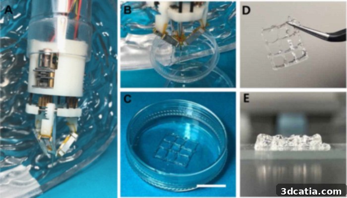
To rigorously test their invention, the Chinese research team ingeniously attached the micro-robot to a standard endoscope. This integrated system was then carefully introduced into a transparent stomach model, allowing for clear observation of its functionality. Upon successfully reaching the target area within the simulated stomach, the prototype initiated its primary function: the precise bioprinting of specialized gels. These gels were meticulously loaded with both epithelial and muscle cells, crucial components for the regeneration of damaged stomach tissue. The ability to deposit these regenerative materials directly onto the stomach wall represents a monumental leap forward in treating internal wounds. This targeted approach promises enhanced healing compared to systemic or less precise methods.
Unprecedented Healing Potential and Future Outlook
The results of these initial experiments were remarkably encouraging. The 3D printed tissues not only maintained their viability but also demonstrated significant proliferation over a period of 10 days. This sustained cellular activity is a critical indicator of the technology’s potential to facilitate the genuine healing of ulcers within the digestive system. Currently, conventional treatments for gastric ulcers can often be protracted and do not always guarantee complete effectiveness, sometimes leaving patients susceptible to recurrence or requiring long-term medication. In contrast, this innovative *in situ* bioprinting project offers a transformative path, potentially representing a substantial advancement in the treatment and management of gastric wounds. It bypasses many of the limitations associated with traditional methods by directly applying regenerative cells to the site of injury.
Looking ahead, Tao Xu, one of the principal authors of the study, shared an ambitious vision for the future evolution of this technology. He states, “Future research could reduce the width of the micro-robot to 12 millimeters and equip it with cameras and other sensors that would enable it to perform more complex operations.” A smaller robot would allow for even less invasive procedures, while integrated cameras would provide real-time visualization, enhancing precision and enabling surgeons to monitor the printing process directly. Additional sensors could potentially gather diagnostic data, assess tissue health, or even assist in navigating more intricate anatomical structures. This future iteration could pave the way for a single device capable of diagnosis, precise wound assessment, and targeted regenerative treatment, offering an unparalleled level of medical intervention.
Overcoming Challenges: Biomaterial Stability and Clinical Translation
Despite the immense promise, the research team encountered a significant challenge during their initial experiments: the stability of the biomaterial. The gels used for bioprinting proved to be stable and maintained their structural integrity only at colder temperatures. At normal human body temperature, however, the material became too liquid, rendering it unsuitable for forming the intricate and stable structures required for effective tissue regeneration. This problem underscored a crucial hurdle for the clinical translation of the technology, as maintaining non-physiological temperatures internally is not feasible.
Addressing this critical issue, another independent study, spearheaded by David Hoelzle and his collaborators, focused on developing alternative biomaterials. Their research successfully identified and developed a new gel formulation that maintained its shape and structural integrity even at warmer, physiological body temperatures. This breakthrough in biomaterial science is vital, as it resolves one of the primary technical barriers to implementing *in situ* bioprinting inside the human body. Hoelzle emphasized the immediate applications of this advanced technique. According to him, while this particular method may not yet be capable of 3D printing complex, fully functional organs with intricate vascularization and innervation, it holds immense potential for significantly enhancing surgical interventions. By enabling the creation of simpler biological structures directly at the site of injury, bioprinting could revolutionize the repair of tissues like gastric lining, skin, or cartilage, thereby accelerating recovery and improving patient outcomes. This focused application allows for practical implementation even as the broader field of organ bioprinting continues its development. For those interested in delving deeper into the specifics of this groundbreaking project, comprehensive information can be found HERE.
The Broader Impact of In Situ Bioprinting in Regenerative Medicine
The development of direct stomach bioprinting represents more than just a new treatment for ulcers; it signifies a pivotal moment for the field of regenerative medicine. The ability to precisely deposit cells and biomaterials directly onto an internal wound site, without extensive open surgery, opens up a myriad of possibilities. This approach minimizes invasiveness, reduces recovery times, and potentially lowers the risk of complications associated with traditional surgical repairs. Imagine a future where internal injuries, from esophageal tears to intestinal damage, could be mended with cellular precision, promoting natural healing processes from within.
The technology also pushes the boundaries of personalized medicine. By utilizing a patient’s own cells, or carefully matched allogeneic cells, the bioprinted tissues could minimize immune rejection, a common challenge in transplantation. This level of customization ensures that the regenerative process is optimally tailored to the individual, leading to more effective and durable repairs. While the path to widespread clinical adoption will involve rigorous testing, regulatory approvals, and further technological refinement, the initial successes of the Tsinghua University and Hoelzle’s teams lay a strong foundation for a future where robotic bioprinters become a standard tool in gastroenterology and internal medicine. The implications extend to areas such as drug delivery, where custom-printed patches could slowly release medication directly at a diseased site, or even for *in vivo* disease modeling to better understand pathological processes in their native environment.
Do you believe that direct bioprinting onto the stomach represents a more effective and less invasive approach to managing gastric problems compared to existing treatments? We invite you to share your thoughts and perspectives in the comments section below, or engage with us on our Facebook and Twitter pages! Don’t miss out on the latest innovations and breakthroughs in the world of 3D printing; sign up for our free weekly Newsletter to receive all the crucial updates directly in your inbox!