
Revolutionizing Bypass Surgery: The Promise of 3D Printed Blood Vessels from Edinburgh
Cardiovascular disease (CVD) stands as the foremost cause of death across the globe, tragically responsible for nearly a third of all fatalities worldwide. This alarming statistic is particularly pronounced in nations such as the USA, where the Centers for Disease Control and Prevention (CDC) reports a death every 33 seconds linked to cardiovascular disease, and in the UK, where heart and circulatory ailments contribute to almost a quarter of all deaths. Given the profound and widespread impact of this health crisis, the continuous pursuit of advanced treatment options is an urgent global priority. In a significant stride toward this goal, researchers at the University of Edinburgh appear to have unearthed a transformative solution, particularly in the realm of coronary artery bypass surgeries. By successfully developing innovative 3D printed blood vessels, their pioneering work holds the potential to fundamentally reshape the landscape of cardiovascular disease treatment, offering new hope to millions of patients.
Understanding Coronary Artery Bypass Surgery and Its Current Challenges
Coronary artery bypass grafting (CABG) is a vital surgical procedure designed to treat advanced coronary artery disease. This serious condition develops when the major arteries supplying oxygen-rich blood to the heart muscle become narrowed or blocked by atherosclerotic plaque. Such blockages can severely restrict blood flow, leading to chest pain (angina), shortness of breath, and in severe cases, heart attacks. CABG aims to circumvent these obstructed arteries by creating new pathways for blood to flow, thereby restoring adequate circulation to the heart. It is one of the most frequently performed cardiac surgeries globally, with approximately 400,000 procedures conducted annually in the United States and around 20,000 in England, underscoring its critical role in managing severe heart conditions.
Despite its high success rate, often quoted at around 98% for immediate outcomes, CABG is a major surgery that carries significant risks and inherent limitations. The risk of complications can be as high as 20%, encompassing issues such as post-operative bleeding, infection, stroke, kidney dysfunction, and myocardial infarction. A primary contributor to these challenges lies in the current methods used to obtain the necessary graft vessels, which are either harvested from the patient’s own body or, less frequently, are synthetic.
Limitations of Traditional Grafting Materials
Historically, bypass surgeries have relied on two main types of grafts: autologous vessels and synthetic conduits. Autologous grafts, sourced from the patient’s own body, are preferred due to their biological compatibility. The most common choices include the saphenous vein from the leg and the internal mammary artery from the chest. While these natural vessels generally offer excellent long-term patency, their harvesting introduces a secondary surgical site with its own set of drawbacks:
- Donor Site Morbidity: Patients frequently experience considerable pain, swelling, bruising, and permanent scarring at the site where the vessel was removed. This can prolong recovery, cause significant discomfort, and increase the risk of localized infections.
- Risk of Infection: Any additional incision increases the overall risk of surgical site infections, which can be challenging to treat and may lead to prolonged hospitalization.
- Nerve Damage: The process of harvesting vessels can sometimes damage surrounding nerves, leading to persistent numbness, tingling, or chronic pain in the donor limb.
- Limited Availability: Not all patients possess healthy and adequate vessels suitable for grafting, especially those with pre-existing vascular conditions, previous surgeries, or advanced age. This can complicate surgical planning and outcomes.
Synthetic grafts, typically manufactured from materials like expanded polytetrafluoroethylene (ePTFE) or woven polyester (Dacron), provide an alternative that bypasses the need for donor site surgery. However, these materials have significant limitations, particularly when used for small-diameter vessels, such as those typically required in coronary bypasses (usually less than 6mm). Synthetic grafts in small-diameter applications often suffer from high failure rates due to:
- Thrombosis: The foreign surface of synthetic materials can trigger blood clot formation, leading to rapid graft occlusion.
- Intimal Hyperplasia: An overgrowth of cells within the vessel wall, leading to gradual narrowing and re-blockage of the graft over time.
- Poor Integration: Synthetic materials struggle to integrate biologically with the surrounding native tissues, leading to a higher risk of infection and lack of long-term stability.
These inherent challenges highlight the urgent need for a superior alternative – a need that the University of Edinburgh’s pioneering development of 3D printed blood vessels aims to address directly.
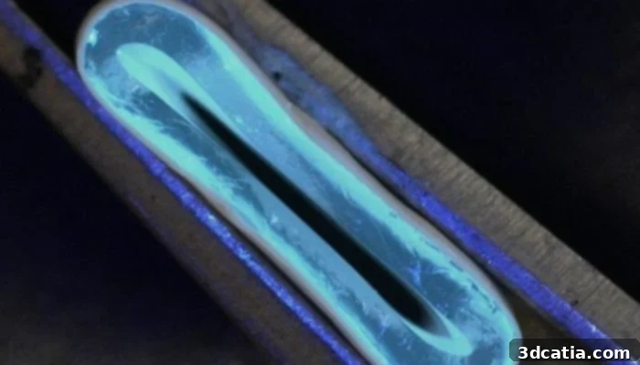
The innovative 3D printed blood vessels (photo credits: Dr. Norbert Radasci, School of Engineering, University of Edinburgh)
The Breakthrough: Engineering 3D Printed Vascular Grafts with Advanced Bioprinting
The innovative concept driving this groundbreaking project is the fabrication of robust, highly flexible, and biologically compatible tubular grafts using sophisticated 3D printing technologies. These engineered blood vessels are specifically designed to serve as direct replacements for both autologous human veins and current synthetic grafts used in bypass operations. This approach seeks to eliminate the significant drawbacks associated with traditional methods, thereby dramatically enhancing patient outcomes, reducing post-operative complications, and streamlining the surgical process.
The advantages offered by these state-of-the-art 3D printed alternatives are extensive and transformative. Firstly, the paramount benefit is the complete elimination of the need for harvesting a human vein from the patient. This instantly removes the associated surgical pain, discomfort, scarring, and the elevated risk of infection at a secondary donor site, leading to a faster, less painful recovery for patients and a less invasive surgical experience. Secondly, these bio-engineered grafts are meticulously designed to surmount the historically high failure rates observed with small-diameter synthetic grafts. The precise control over material composition, structural architecture, and mechanical properties afforded by 3D printing ensures that these artificial vessels can integrate more effectively and durably into the human circulatory system. This meticulous design reduces the likelihood of complications such as thrombosis (blood clot formation) and intimal hyperplasia (thickening of the vessel wall), which often lead to the re-occlusion of traditional synthetic grafts.
The Innovative Two-Stage Fabrication Process: A Deeper Dive
To achieve these ambitious and potentially life-saving outcomes, the University of Edinburgh team pioneered a novel and highly sophisticated two-stage fabrication process. This ingenious method seamlessly integrates principles of advanced additive manufacturing with cutting-edge material science, ensuring the production of grafts that possess the critical mechanical strength, elasticity, and biological compatibility required to function flawlessly within the dynamic environment of the human body.
The initial and foundational step involves an innovative application of extrusion-based 3D printing. At the core of this stage is a precisely controlled rotating spindle, carefully integrated into a specialized 3D printer setup. The team utilizes this sophisticated system to extrude tubular grafts using a specifically formulated water-based gel, often referred to in bioprinting as a ‘bio-ink.’ The rotating spindle plays an indispensable role by ensuring the uniform thickness, consistent diameter, and overall structural integrity of the nascent tubular scaffold, effectively mimicking the natural cylindrical architecture of blood vessels. The choice of a water-based gel is strategic; it serves as a temporary, biocompatible scaffold, providing the initial structural framework while being gentle enough to potentially allow for future cellular integration and tissue regeneration, setting the stage for true vascular tissue engineering.
Following the precise extrusion phase, the newly formed grafts undergo a crucial reinforcement process: electrospinning. This advanced technique is pivotal in endowing the artificial blood vessels with the necessary mechanical strength, resilience, and flexibility. During electrospinning, a high voltage is applied to a polymer solution, which then draws out incredibly fine, continuous nanofibers. These nanofibers are meticulously deposited onto the surface of the extruded tubular graft, creating a dense, interwoven mesh-like coating. The material selected for this vital reinforcement layer is a biodegradable polyester. This choice is deliberate and optimized, as biodegradable polyesters are celebrated for their exceptional biocompatibility, their ability to degrade predictably over time, and their tunable mechanical properties, which can be engineered to closely match those of natural biological tissues. The resulting nanofiber coating significantly enhances the graft’s biomechanical properties, providing the required elasticity and long-term durability to withstand the constant pulsatile pressures of the circulatory system, while simultaneously offering a conducive microenvironment and scaffold for the patient’s own cells to colonize and eventually remodel the graft into living tissue.
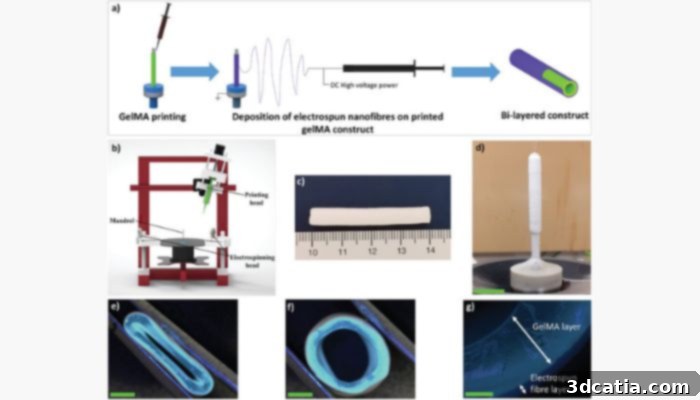
Detailed diagram showing the innovative process for creating the 3D printed blood vessels (Photo Credits: Dr. Norbert Radasci, School of Engineering, University of Edinburgh)
Achieved Results and the Promising Path to Clinical Application
The meticulous development and comprehensive testing of these innovative grafts have yielded exceptionally promising results, marking a significant milestone in vascular tissue engineering. The University of Edinburgh team has successfully engineered 3D printed blood vessels that can be precisely manufactured across an impressive and versatile range of diameters, spanning from a minute 1 mm up to a substantial 40 mm. This broad spectrum of sizes is immensely critical, as it ensures the potential for creating grafts suitable for diverse applications within the complex cardiovascular system, from the smallest coronary arteries to larger peripheral vessels, thereby addressing a wider array of clinical needs. Furthermore, a pivotal achievement lies in the remarkable flexibility of these printed vessels. This inherent pliability makes them considerably easier to manipulate and integrate into the intricate and dynamic anatomical environment of the human body during surgery, minimizing trauma during implantation and promoting superior long-term function and patency.
Crucially, extensive mechanical evaluations have unequivocally demonstrated that these bio-engineered products possess both strength and elasticity that are remarkably comparable to those of natural human blood vessels. This biomechanical fidelity is paramount for successful and durable implantation, as it ensures the grafts can reliably withstand the constant physiological blood pressures and pulsatile flow without succumbing to rupture, fatigue, or premature degradation. The successful replication of these critical natural properties represents a monumental leap forward in the field of vascular tissue engineering, effectively resolving a long-standing challenge in the creation of functional and enduring artificial blood vessels.
Future Steps: Navigating the Journey from Laboratory to Patient Care
While the laboratory results are profoundly encouraging, the journey from this innovative discovery to widespread clinical application involves a series of carefully planned and rigorously executed steps. The immediate next phase of this vital research will concentrate on comprehensive in-vivo testing of these 3D printed blood vessels within relevant animal models. These critical studies are indispensable for evaluating the grafts’ long-term patency (remaining open), biocompatibility (how well they interact with living tissue), and their successful integration within a living biological system. Researchers will meticulously monitor for any signs of immune rejection, thrombosis, infection, and critically observe how effectively the grafts become vascularized and integrate with the surrounding native tissues. This pivotal phase will furnish invaluable data regarding the safety, efficacy, and long-term performance of the grafts, which is absolutely essential before proceeding to human clinical trials.
Should the animal trials demonstrate unequivocal success, the research will then advance to the rigorous stages of human clinical trials. This is a highly regulated and multi-phase process, commencing with small groups of patients to primarily assess safety, followed by larger, more extensive trials to robustly determine efficacy when compared to existing standard treatments. The ultimate and ambitious vision is to secure comprehensive regulatory approval from health authorities worldwide, thereby paving the way for these advanced 3D printed blood vessels to become a standard, readily available treatment option. Such a development could potentially transform cardiovascular care, allowing for “off-the-shelf” solutions that would significantly reduce surgical waiting times and dramatically improve access to life-saving procedures for countless patients suffering from cardiovascular disease globally.
Dr. Norbert Radacsi, the esteemed principal investigator from the University of Edinburgh’s School of Engineering, eloquently articulated the profound significance of this scientific achievement. He stated, “The results from our research address a long-standing challenge in the field of vascular tissue engineering – to produce a conduit that has similar biomechanical properties to that of human veins. With continued support and collaboration, the vision of improved treatment options for patients with cardiovascular disease could become a reality.” His insightful statement perfectly encapsulates the immense potential of this groundbreaking work to usher in a new, transformative era in cardiovascular care and regenerative medicine. For those interested in delving deeper, more detailed scientific findings are comprehensively available in the full study, which can be accessed HERE.
Broader Implications and the Future Landscape of Regenerative Medicine
The implications of this significant breakthrough extend far beyond the immediate application in coronary artery bypass surgery. The successful development of customizable, mechanically robust, and highly biocompatible 3D printed blood vessels unlocks a myriad of possibilities for numerous other applications within vascular surgery, tissue engineering, and the broader field of regenerative medicine. This innovative technology could be readily adapted for repairing a wide range of damaged or diseased blood vessels throughout the entire body, offering novel therapeutic avenues for conditions such as peripheral artery disease, which affects millions. Furthermore, this capability to engineer vascular conduits is crucial for creating intricate vascular networks necessary for the successful regeneration of complex tissues and even entire organs, pushing the boundaries of what is medically possible.
Pediatric cardiovascular surgery, a field often fraught with unique challenges due to the small size of vessels and the critical need for grafts that can grow and adapt with the child, stands to benefit immensely from this innovative approach. The unparalleled ability to precisely tailor graft dimensions, mechanical properties, and biological characteristics for individual patients heralds a new, exciting era of truly personalized medicine in cardiovascular care, moving away from a one-size-fits-all approach. This pioneering research by the University of Edinburgh serves as a powerful testament to the transformative power of additive manufacturing and advanced bioprinting in revolutionizing healthcare. By seamlessly combining engineering ingenuity with profound medical understanding, scientists are now capable of creating sophisticated biological substitutes that were once relegated to the realm of science fiction. As 3D printing technology continues its rapid advancement and refinement, we can confidently anticipate further innovations that will not only substantially improve existing treatments but also unlock entirely new therapeutic possibilities for a vast array of diseases. The successful development of these 3D printed blood vessels is more than just a scientific achievement; it is a beacon of hope, promising to significantly alleviate the global burden of cardiovascular disease and dramatically enhance the quality of life for millions of patients around the world.
What are your thoughts on these revolutionary 3D printed blood vessels and their potential impact on cardiovascular health? We eagerly encourage you to share your valuable insights and comments below, or engage with us and join the conversation on our vibrant social media platforms: LinkedIn, Facebook, and Twitter pages! To ensure you never miss out on the very latest advancements and breaking news in 3D printing and medical technology, be sure to sign up for our free weekly newsletter here, delivering top stories directly to your inbox. Furthermore, you can explore our extensive and insightful video library on our dedicated YouTube channel for more in-depth content and visual demonstrations.