
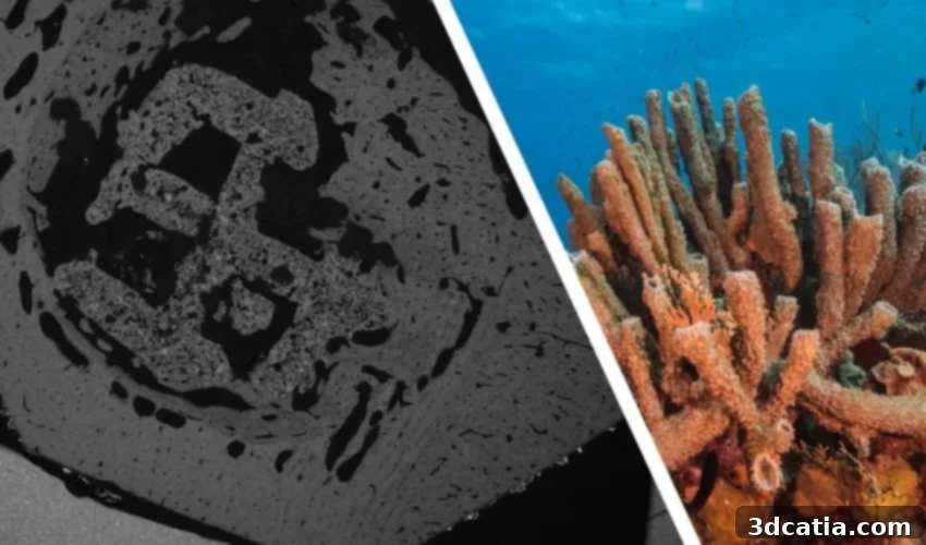
The Future of Bone Repair: Swansea University’s 3D Printed Coral-Inspired Grafts Offer Revolutionary Healing
A groundbreaking discovery at Swansea University is set to transform regenerative medicine, specifically in the field of bone repair. A dedicated team of researchers has successfully 3D printed a novel bone graft material that draws its inspiration from the intricate structure of coral. What makes this innovation particularly remarkable is its dual benefit: the new material not only facilitates significantly faster bone healing but also undergoes a natural degradation process within the body once the repair is complete, leaving behind healthy, regenerated bone tissue. This breakthrough addresses critical limitations of current bone grafting techniques, paving the way for more effective and natural healing processes for patients worldwide.
Bone defects, resulting from a myriad of causes such as severe fractures, tumor resections, or non-healing injuries, represent a substantial global health challenge and are among the leading causes of long-term disability. While bone tissue possesses an inherent capacity for self-repair and regeneration, this natural healing process can be severely compromised or slowed down by various factors, including the patient’s age, underlying diseases, or the sheer size and complexity of the defect. For large or critical-sized bone defects, the body often requires external assistance in the form of a scaffold or a bone graft to bridge the gap and provide a framework for new bone growth, thereby supplementing and accelerating tissue regeneration.
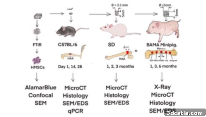
Design of in vitro and in vivo tests (Credits: Swansea University)
Historically, medical professionals have relied on autografts, which involve transplanting bone from another part of the patient’s own body, or allografts, which utilize bone from a donor. While effective in certain scenarios, both traditional methods come with significant drawbacks. Autografts are limited by the availability of donor bone, can cause considerable pain and complications at the donor site (known as donor site morbidity), and often require additional surgical procedures. Allografts, on the other hand, face issues such as limited supply, the potential for immune rejection, the risk of disease transmission, and ethical considerations surrounding donor tissue. These challenges underscore the urgent need for safe, effective, and readily available alternatives for bone regeneration.
Existing synthetic bone grafts have attempted to address these limitations but have largely failed to replicate the intricate performance and biological integration of natural bone. Many synthetic options degrade too slowly, leading to persistent foreign material in the body, or fail to integrate effectively with surrounding native tissue. Some can even trigger adverse side effects, such as chronic inflammation or mechanical incompatibility, ultimately hindering optimal healing. In stark contrast, the innovative material developed by the Swansea team represents a paradigm shift. It is meticulously engineered to closely mimic the structural characteristics and biological behavior of natural bone, effectively overcoming the multifaceted challenges associated with conventional and existing synthetic graft alternatives. Its design fosters harmonious integration with the body’s natural healing processes, promising superior patient outcomes.
Why Coral’s Unique Structure Offers a Solution
The exploration of corals as potential bone graft substitutes began as early as the 1970s, driven by the compelling observation that certain coral species exhibit striking similarities to cancellous bone, also known as spongy bone. This natural resemblance extends beyond mere appearance, encompassing crucial mechanical properties and an inherent biocompatibility. Corals are naturally porous calcium carbonate structures, and their interconnected pore network provides an ideal scaffold for cellular infiltration and nutrient exchange, essential elements for new bone formation. Furthermore, corals are osteoconductive, meaning they can guide the growth of new bone tissue, and biodegradable, allowing for their gradual resorption as the host bone regenerates.
Over decades, extensive research, including findings published in the Journal of International Oral Health, has consistently affirmed natural coral porous calcium carbonate as a “clinically useful bone replacement graft material.” Building upon this rich history and understanding, the Swansea team embarked on developing a sophisticated new material. Their innovation involves mimicking both the highly porous architecture and the precise chemical composition of a coral-converted bone graft substitute. This biomimetic approach ensures that the synthetic graft behaves as similarly to natural bone as possible. The material was then precisely engineered using advanced 3D printing techniques, specifically a 3D-Bioplotter from Envisiontec, to create custom-shaped implants. Alternatively, the material could also be molded and left to dry at room temperature, offering versatility in manufacturing. This method allows for the creation of intricate, patient-specific scaffolds that perfectly integrate into the defect site.
To rigorously test the efficacy and safety of this novel material, the research team conducted comprehensive preclinical in vivo trials, utilizing animal models including rats and minipigs. The results from these trials were nothing short of remarkable. They conclusively demonstrated that the coral-inspired material was capable of fully repairing bone defects within an impressive timeframe of three to six months. Even more significantly, the material actively triggered and supported the formation of a robust new layer of strong, healthy cortical bone – the dense outer layer of bone – in as little as four weeks. This rapid regeneration and formation of high-quality bone tissue represent a critical advancement over existing bone graft technologies, promising significantly faster patient recovery and improved functional outcomes.
Revolutionary Outcomes and Global Impact
The uniquely engineered structure of this 3D printed bone graft offers a multitude of incredible benefits that stand to revolutionize bone repair. Foremost among these is the unprecedented speed of healing; new bone growth was observed to occur in just two to four weeks post-implantation. This dramatically reduces the patient’s recovery period and minimizes the risks associated with prolonged healing. Furthermore, the material exhibits a crucial property of natural degradation. Within six to twelve months after successful bone regeneration, the graft naturally dissolves and is absorbed by the body, leaving behind only the patient’s own healthy, newly formed bone. This eliminates the need for removal surgeries and prevents the long-term presence of foreign materials, which can often lead to complications or stress shielding in traditional implants.
Beyond its biological advantages, the material also boasts significant logistical and economic benefits. Unlike its traditional alternatives, such as natural coral or donor bone, this synthetic, 3D printed alternative is far easier to access and produce. Its scalable manufacturing process allows for the creation of large quantities, ensuring a consistent and reliable supply. This widespread availability stands to dramatically reduce the existing reliance on limited donor bone resources, thereby mitigating ethical concerns and alleviating supply chain issues that often plague current bone grafting practices. The ability to produce these grafts on demand and in tailored shapes also opens doors for personalized medicine, where implants can be customized to precisely fit a patient’s unique defect.
The pioneering research was spearheaded by Dr. Zhidao Xia of Swansea University Medical School, in collaboration with esteemed faculty members from the university’s Science and Engineering departments, as well as vital external partners. The team officially published their patented research findings late this November, marking a significant milestone in regenerative medicine and biomedical engineering.
“Our invention bridges the critical gap between less effective synthetic substitutes and the gold standard of donor bone,” Dr. Xia articulated. “We have definitively demonstrated that it is possible to create a material that is not only safe and highly effective but also scalable to meet the immense global demand for bone grafts. This breakthrough holds the potential to effectively end the reliance on donor bone, simultaneously addressing the complex ethical dilemmas and pervasive supply chain challenges inherent in traditional bone grafting procedures.”
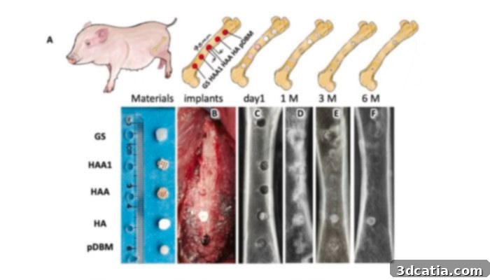
Implants in minipig (Credits: Swansea University)
Building on this remarkable success, the Swansea University team is actively seeking collaborations with leading companies and healthcare organizations to make their innovative technology globally accessible. This 3D printed coral-inspired bone graft alternative promises significant societal benefits, including the potential to considerably reduce healthcare costs associated with bone repair surgeries and, most importantly, to dramatically improve the quality of life for countless patients suffering from bone defects. The impact of this research extends far beyond immediate patient care; it opens up unprecedented new avenues and exciting opportunities for further innovation within the broader biomedical industry, fostering advancements in regenerative therapies and personalized medicine. To delve deeper into this transformative research and its implications, interested parties can read Swansea University’s official press release here.
What are your thoughts on these pioneering 3D printed coral-inspired bone grafts and their potential to revolutionize bone repair? Let us know in a comment below or connect with us on ourLinkedIn,Facebook, andTwitter pages! Don’t miss out on the latest advancements in additive manufacturing – be sure to sign up for our free weeklyNewsletter here, delivering the most relevant 3D printing news straight to your inbox! You can also explore all our insightful videos on ourYouTube channel.
*Cover Photo Credits: (Left) An image of a 3D-printed material implanted in vivo for 4 weeks, captured using a scanning electron microscope. Credit: Dr. Zhidao Xia. (Right) A vibrant photo of natural coral. Credit: Jesus Cobaleda.